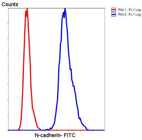
免费电话:400-9961-933
邮箱:techcn@signalwayantibody.com
欢迎光临本站
登录 注册 English
位置: 首页 > 抗体 > N-Cadherin Antibody

N-Cadherin Antibody#48495

N-Cadherin Antibody
N-Cadherin Antibody
N-Cadherin Antibody
N-Cadherin Antibody
N-Cadherin Antibody
N-Cadherin Antibody
N-Cadherin Antibody
是否有货: Yes
产品总价:
产品详情

产品名称N-Cadherin Antibody

来源种属Rabbit

克隆性Polyclonal

纯化Peptide affinity purified

应用WB, ICC, IHC, FC

种属反应性Hu, Ms

免疫原描述Synthetic peptide (KLH-coupled) within human N-cadherin 360-420 aa.

别名CADH2_HUMAN antibody
Cadherin 2 antibody
Cadherin 2 N cadherin neuronal antibody
Cadherin 2 type 1 antibody
Cadherin 2 type 1 N cadherin neuronal antibody
Cadherin 2, type 1, N-cadherin (neuronal) antibody
Cadherin-2 antibody
Cadherin2 antibody
Calcium dependent adhesion protein neuronal antibody
CD325 antibody
CD325 antigen antibody
CDH2 antibody
CDHN antibody
CDw325 antibody
CDw325 antigen antibody
N cadherin 1 antibody
N-cadherin antibody
NCAD antibody
Neural cadherin antibody
OTTHUMP00000066304 antibody
OTTHUMP00000067378 antibody

数据库入口号Swiss-Prot#:P19022

计算分子量140 kDa

配方1*TBS (pH7.4), 1%BSA, 40%Glycerol. Preservative: 0.05% Sodium Azide.

保存Store at -20˚C

应用详情
WB: 1:500
IHC: 1:200
ICC: 1:200

FC: 1:50
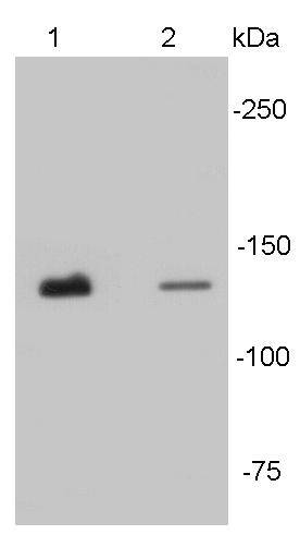
Western blot analysis of N-cadherin on different tissue lysates using anti- N-cadherin antibody at 1/500 dilution. Positive control:
Lane 1: Mouse heart
Lane2 : Human heart
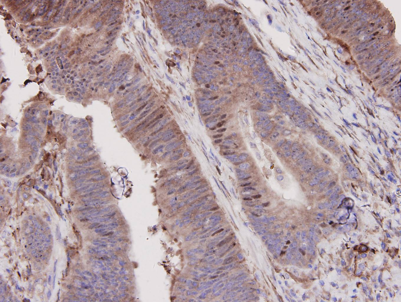
Immunohistochemical analysis of paraffin-embedded human colon carcinoma tissue using anti-N-Cadherin antibody. Counter stained with hematoxylin.
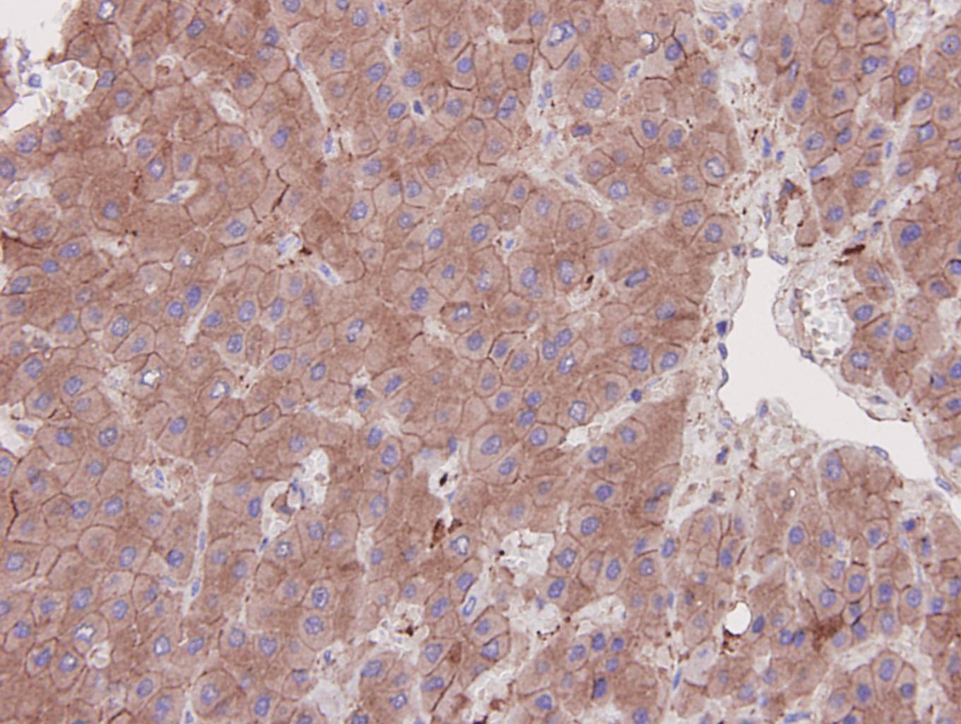
Immunohistochemical analysis of paraffin-embedded human liver carcinoma tissue using anti-N-Cadherin antibody. Counter stained with hematoxylin.
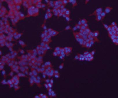
ICC staining N-Cadherin in 293 cells (red). The nuclear counter stain is DAPI (blue). Cells were Cells were fixed in paraformaldehyde, permeabilised with 0.25% Triton X100/PBS.
ICC staining N-Cadherin in mouse embryonic stem cells (green). The nuclear counter stain is DAPI (blue). Cells were Cells were fixed in paraformaldehyde, permeabilised with 0.25% Triton X100/PBS.
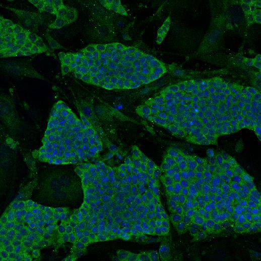
ICC staining N-Cadherin in HepG2 cells (green). The nuclear counter stain is DAPI (blue). Cells were Cells were fixed in paraformaldehyde, permeabilised with 0.25% Triton X100/PBS.
背景
The cadherins are a large family of cell adhesion molecules involved in calcium-dependent recognition and adhesion events. The cadherin subfamily includes N-, P-, B-, and E- cadherins, and other members. Recent studies indicate that cancer cells have up-regulated N-cadherin in addition to loss of E-cadherin. N-cadherin cooperates with the FGF receptor, leading to overexpression of MMP-9 and cellular invasion. N-Cadherin is commonly found in cancer cells and provides a mechanism for transendothelial migration. When a cancer cell adheres to the endothelial cells of a blood vessel it up-regulates the src kinase pathway, which phosphorylates beta-catenins attached to both N-cadherin (this protein) and E-cadherins.
背景文献
1. Liu J et al. Loss of DLG5 promotes breast cancer malignancy by inhibiting the Hippo signaling pathway. Sci Rep 7:42125-42125 (2017).
2. Chen R et al. Glycoproteomics analysis of human liver tissue by combination of multiple enzyme digestion and hydrazide chemistry. J Proteome Res 8:651-661 (2009).

如果您使用该产品48495发表了文章,请通知我们,让我们可以引用您的文献。

et al,Neferine, a novel ROCK1-targeting inhibitor, blocks EMT process and induces apoptosis in non-small cell lung cancer. In J Cancer Res Clin Oncol on 2022 Aug 19 by Po Hu, Peng Wan, et al..PMID:35984492, , (2022),
PMID: 35984492
5

注释

应用

  • WB免疫印迹
  • IHC免疫组化
  • IF免疫荧光
  • ICC免疫细胞化学
  • FC流式细胞
  • IP免疫沉淀
  • E酶联免疫吸附法
  • DB免疫斑点法
  • ChIP染色质免疫沉淀
  • GICA胶体金免疫层析法
  • NC阴性对照

种属反应性

  • Hu
  • Ms小鼠
  • Rt大鼠
  • Dm果蝇
  • C线虫
  • Mk
  • Rb
  • B
  • D
  • P
  • Hm仓鼠
  • ChHm中国仓鼠
  • Chk

联系我们

Location :

南京市高新开发区星火路10号人才大厦C座

Phone :

400-9961-933
025-58868422

Email Address

techcn@signalwayantibody.com