免费电话:400-9961-933
邮箱:techcn@signalwayantibody.com
欢迎光临本站
登录 注册 English
位置: 首页 > 重组兔单抗 > PI 3 Kinase p85 alpha Rabbit mAb

PI 3 Kinase p85 alpha Rabbit mAb#48848

评价
Recombinant
PI 3 Kinase p85 alpha Rabbit mAb
是否有货: Yes
产品总价:
产品详情

产品名称PI 3 Kinase p85 alpha Rabbit mAb

克隆性SU04-07

纯化ProA affinity purified

应用WB, ICC/IF, IHC, FC

种属反应性Hu, Ms, Rt

免疫原描述Synthetic peptide within C-terminal human PI 3 Kinase p85 alpha.

别名GRB1 antibody
p85 alpha antibody
p85 antibody
P85A_HUMAN antibody
Phosphatidylinositol 3 kinase associated p 85 alpha antibody
Phosphatidylinositol 3 kinase regulatory 1 antibody
Phosphatidylinositol 3 kinase, regulatory subunit, polypeptide 1 (p85 alpha) antibody
Phosphatidylinositol 3-kinase 85 kDa regulatory subunit alpha antibody
Phosphatidylinositol 3-kinase regulatory subunit alpha antibody
Phosphoinositide 3 kinase, regulatory subunit 1 (alpha) antibody
PI3 kinase p85 subunit alpha antibody
PI3-kinase regulatory subunit alpha antibody
PI3-kinase subunit p85-alpha antibody
PI3K antibody
PI3K regulatory subunit alpha antibody
Pik3r1 antibody
PtdIns 3 kinase p85 alpha antibody
PtdIns-3-kinase regulatory subunit alpha antibody
PtdIns-3-kinase regulatory subunit p85-alpha antibody

数据库入口号Swiss-Prot#:P27986

计算分子量84 kDa

浓度1 mg/mL

配方1*TBS (pH7.4), 0.05% BSA, 40% Glycerol. Preservative: 0.05% Sodium Azide.

保存Store at -20˚C

应用详情

WB: 1:1,000-1:2,000
IHC: 1:50-1:200
ICC: 1:50-1:200

FC: 1:50-1:100

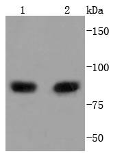
Western blot analysis of PI 3 Kinase p85 alpha on different lysates using anti-PI 3 Kinase p85 alpha antibody at 1/1,000 dilution. Positive control:
Lane 1: MCF-7
Lane 2: Raji
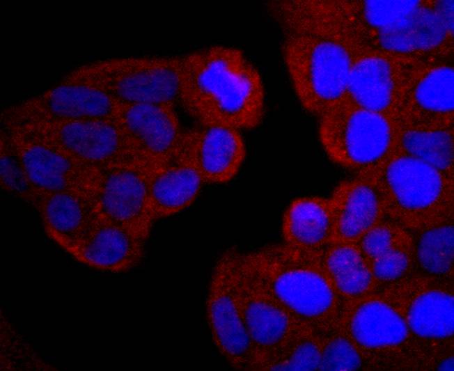
ICC staining PI 3 Kinase p85 alpha in Hela cells (red). The nuclear counter stain is DAPI (blue). Cells were fixed in paraformaldehyde, permeabilised with 0.25% Triton X100/PBS.
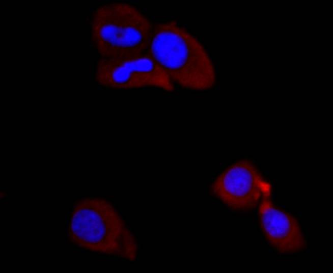
ICC staining PI 3 Kinase p85 alpha in MCF-7 cells (red). The nuclear counter stain is DAPI (blue). Cells were fixed in paraformaldehyde, permeabilised with 0.25% Triton X100/PBS.
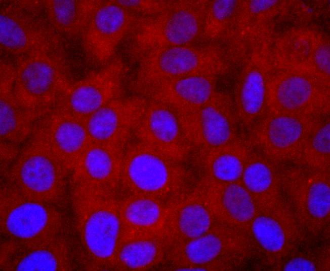
ICC staining PI 3 Kinase p85 alpha in HepG2 cells (red). The nuclear counter stain is DAPI (blue). Cells were fixed in paraformaldehyde, permeabilised with 0.25% Triton X100/PBS.
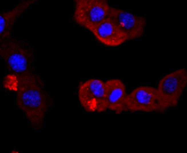
ICC staining PI 3 Kinase p85 alpha in NIH/3T3 cells (red). The nuclear counter stain is DAPI (blue). Cells were fixed in paraformaldehyde, permeabilised with 0.25% Triton X100/PBS.
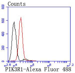
Flow cytometric analysis of HepG2 cells with PI 3 Kinase p85 alpha antibody at 1/50 dilution (red) compared with an unlabelled control (cells without incubation with primary antibody; black). Alexa Fluor 488-conjugated goat anti rabbit IgG was used as the secondary antibody
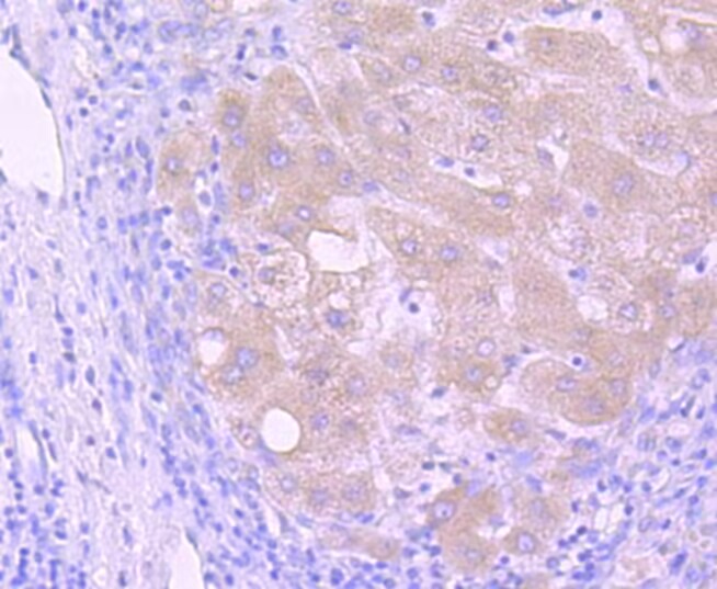
Immunohistochemical analysis of paraffin-embedded human liver carcinoma tissue using anti-PI 3 Kinase p85 alpha antibody. The section was pre-treated using heat mediated antigen retrieval with Tris-EDTA buffer (pH 8.0-8.4) for 20 minutes.The tissues were blocked in 5% BSA for 30 minutes at room temperature, washed with ddH2O and PBS, and then probed with the primary antibody (1/50) for 30 minutes at room temperature. The detection was performed using an HRP conjugated compact polymer system. DAB was used as the chromogen. Tissues were counterstained with hematoxylin and mounted with DPX.
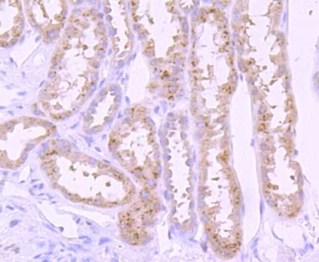
Immunohistochemical analysis of paraffin-embedded human kidney tissue using anti-PI 3 Kinase p85 alpha antibody. The section was pre-treated using heat mediated antigen retrieval with Tris-EDTA buffer (pH 8.0-8.4) for 20 minutes.The tissues were blocked in 5% BSA for 30 minutes at room temperature, washed with ddH2O and PBS, and then probed with the primary antibody (1/50) for 30 minutes at room temperature. The detection was performed using an HRP conjugated compact polymer system. DAB was used as the chromogen. Tissues were counterstained with hematoxylin and mounted with DPX
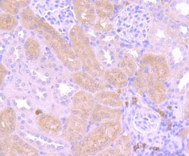
Immunohistochemical analysis of paraffin-embedded mouse kidney tissue using anti-PI 3 Kinase p85 alpha antibody. The section was pre-treated using heat mediated antigen retrieval with Tris-EDTA buffer (pH 8.0-8.4) for 20 minutes.The tissues were blocked in 5% BSA for 30 minutes at room temperature, washed with ddH2O and PBS, and then probed with the primary antibody (1/50) for 30 minutes at room temperature. The detection was performed using an HRP conjugated compact polymer system. DAB was used as the chromogen. Tissues were counterstained with hematoxylin and mounted with DPX.
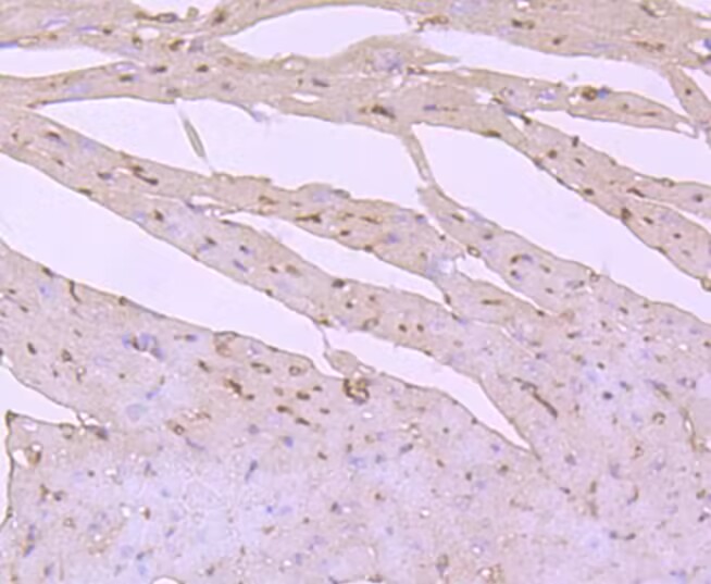
Immunohistochemical analysis of paraffin-embedded mouse heart tissue using anti-PI 3 Kinase p85 alpha antibody. The section was pre-treated using heat mediated antigen retrieval with Tris-EDTA buffer (pH 8.0-8.4) for 20 minutes.The tissues were blocked in 5% BSA for 30 minutes at room temperature, washed with ddH2O and PBS, and then probed with the primary antibody (1/50) for 30 minutes at room temperature. The detection was performed using an HRP conjugated compact polymer system. DAB was used as the chromogen. Tissues were counterstained with hematoxylin and mounted with DPX.
背景

Phosphatidylinositol 3-kinase (PI 3-kinase) phosphorylates the 3' OH position of the inositol ring of inositol lipids and is composed of p85 and p110 subunits. PI 3-kinase p85 lacks PI 3-kinase activity and acts as an adapter, coupling p110 to activated protein tyrosine kinase. Two forms of p85 have been described (p85α and p85β), each possessing one SH3 and two SH2 domains. PI 3-kinase p85α, also known as GRB1, phosphatidylinositol 3-kinase regulatory 1 or p85, is a 724 amino acid protein that exists as four alternatively spliced isoforms. Involved in insulin metabolism, defects in the PI 3-kinase p85α gene have been linked to insulin resistance. PI 3-kinase p85α is polyubiquitinated in T-cells by Cbl-b, and has multiple phosphorylated amino acid residues, including a phosphorylated tyrosine residue at position 467.

背景文献

1. Yan LX et al. PIK3R1 targeting by miR-21 suppresses tumor cell migration and invasion by reducing PI3K/AKT signaling and reversing EMT, and predicts clinical outcome of breast cancer. Int J Oncol 48:471-84 (2016).
2. Hu J et al. Filamin B regulates chondrocyte proliferation and differentiation through Cdk1 signaling. PLoS One 9:e89352 (2014).

如果您使用该产品48848发表了文章,请通知我们,让我们可以引用您的文献。

et al,Effect of Fushengong Decoction on PTEN/PI3K/AKT/NF-KB Pathway in Rats With Chronic Renal Failure via Dual-Dimension Network Pharmacology Strategy.In Front Pharmacol on 2022 Mar 15 by Hongyu Luo, Munan Wang,et al..PMID: 35370667, , (2022),
PMID: 35370667
et al,Effect of Fushengong Decoction on PTEN/PI3K/AKT/NF-κB Pathway in Rats With Chronic Renal Failure via Dual-Dimension Network Pharmacology Strategy. In Front Pharmacol on 2022 Mar 15 by Hongyu Luo, Munan Wang, et al..PMID: 35370667, , (2022),
PMID: 35370667
5

注释

应用

  • WB免疫印迹
  • IHC免疫组化
  • IF免疫荧光
  • ICC免疫细胞化学
  • FC流式细胞
  • IP免疫沉淀
  • E酶联免疫吸附法
  • DB免疫斑点法
  • ChIP染色质免疫沉淀
  • GICA胶体金免疫层析法
  • NC阴性对照

种属反应性

  • Hu
  • Ms小鼠
  • Rt大鼠
  • Dm果蝇
  • C线虫
  • Mk
  • Rb
  • B
  • D
  • P
  • Hm仓鼠
  • ChHm中国仓鼠
  • Chk

联系我们

Location :

南京市高新开发区星火路10号人才大厦C座

Phone :

400-9961-933
025-58868422

Email Address

techcn@signalwayantibody.com