免费电话:400-9961-933
邮箱:techcn@signalwayantibody.com
欢迎光临本站
登录 注册 English
位置: 首页 > 磷酸化抗体 > NMDAR1 (Phospho-Ser890) Antibody

NMDAR1 (Phospho-Ser890) Antibody#11979

NMDAR1 (Phospho-Ser890) Antibody
是否有货: 有货
产品总价:
产品详情

产品名称NMDAR1 (Phospho-Ser890) Antibody

来源种属Rabbit

克隆性Polyclonal

纯化Antibodies were produced by immunizing rabbits with synthetic phosphopeptide and KLH conjugates. Antibodies were purified by affinity-chromatography using epitope-specific phosphopeptide. Non-phospho specific antibodies were removed by chromatogramphy using non-phosphopeptide.

应用WB, IH, IF/IC

种属反应性Hu Ms Rt

特异性The antibody detects endogenous level of NMDAR1 only when phosphorylated at serine 890.

免疫原类型Peptide-KLH

免疫原描述Peptide sequence around phosphorylation site of serine890(A-S-S(p)-F-K) derived from Human NMDAR1 .

基因/蛋白名称NMDAR1

修饰Phospho

别名GLURZ1; GRIN1; N-methyl-D-aspartate receptor; NMZ1; NMDZ1

数据库入口号Swiss-Prot#: Q05586;
NCBI Gene#: 2902;
NCBI Protein#: NP_000823.4

UniprotQ05586

实际分子量105kd

浓度1.0mg/ml

配方Rabbit IgG in phosphate buffered saline (without Mg2+ and Ca2+), pH 7.4, 150mM NaCl, 0.02% sodium azide and 50% glycerol.

保存Store at -20˚C/1 year

应用详情

Western blotting: 1:500~1:1000

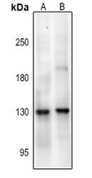
Western blot analysis of NMDAR1 (pS890) expression in A549 (A), U87MG (B) whole cell lysates.
Immunohistochemical analysis of NMDAR1 (pS890) staining in human brain formalin fixed paraffin embedded tissue section. The section was pre-treated using heat mediated antigen retrieval with sodium citrate buffer (pH 6.0). The section was then incubated with the antibody at room temperature and detected using an HRP conjugated compact polymer system.
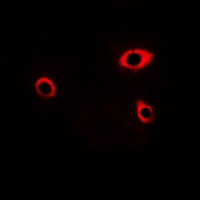
Immunofluorescent analysis of NMDAR1 (pS890) staining in A549 cells. Formalin-fixed cells were permeabilized with 0.1% Triton X-100 in TBS for 5-10 minutes and blocked with 3% BSA-PBS for 30 minutes at room temperature. Cells were probed with the primary antibody in 3% BSA-PBS and incubated overnight at 4 °C in a hidified chamber. Cells were washed with PBST and incubated with a Alexa Fluor 594-conjugated secondary antibody (red) in PBS at room temperature in the dark.
背景

NMDA receptors are members of the ionotropic class of glutamate receptors, which also includes Kainate and AMPA receptors. NMDA receptors consist of NR1 subunits combined with one or more NR2 (A-D) or NR3 (A-B) subunits. The ligand-gated channel is permeable to cations including Ca2+, and at resting membrane potentials NMDA receptors are inactive due to a voltage-dependent blockade of the channel pore by Mg2+. NMDA receptor activation, which requires binding of glutamate and glycine, leads to an influx of Ca2+ into the postsynaptic region where it activates several signaling cascades, including pathways leading to the induction of long-term potentiation (LTP) and depression (LTD). NMDA receptors have a critical role in excitatory synaptic transmission and plasticity in the CNS. They govern a range of physiological conditions including neurological disorders caused by excitotoxic neuronal injury, psychiatric disorders and neuropathic pain syndromes.

Xu M, Smothers CT, Woodward JJ (2011) Alcohol 45, 373-80. Kong M, Ba M, Song L, Liu Z (2009)Neurochem Res 34, 1513-21 . Jackson MF, et al. (2006)J Neurochem 96, 1509-18.

如果您使用该产品11979发表了文章,请通知我们,让我们可以引用您的文献。

5

注释

应用

  • WB免疫印迹
  • IHC免疫组化
  • IF免疫荧光
  • ICC免疫细胞化学
  • FC流式细胞
  • IP免疫沉淀
  • E酶联免疫吸附法
  • DB免疫斑点法
  • ChIP染色质免疫沉淀
  • GICA胶体金免疫层析法
  • NC阴性对照

种属反应性

  • Hu
  • Ms小鼠
  • Rt大鼠
  • Dm果蝇
  • C线虫
  • Mk
  • Rb
  • B
  • D
  • P
  • Hm仓鼠
  • ChHm中国仓鼠
  • Chk

联系我们

Location :

南京市高新开发区星火路10号人才大厦C座

Phone :

400-9961-933
025-58868422

Email Address

techcn@signalwayantibody.com