产品详情
产品名称IRF1 Rabbit mAb
来源种属Recombinant Rabbit
克隆性 Monoclonal antibody
克隆性SR44-08
纯化ProA affinity purified
应用WB, ICC/IF, IHC, IP, FC
种属反应性Hu, Ms, Rt
免疫原描述recombinant protein
别名Interferon regulatory factor 1 antibody
Interferon regulatory factor 1 isoform +I9 antibody
Interferon regulatory factor 1 isoform d78 antibody
Interferon regulatory factor 1 isoform delta4 antibody
Interferon regulatory factor 1 isoform delta7 antibody
IRF 1 antibody
IRF-1 antibody
IRF1 antibody
IRF1_HUMAN antibody
MAR antibody
MAR1 antibody
数据库入口号Swiss-Prot#:P10914
计算分子量48 kDa
配方1*TBS (pH7.4), 1%BSA, 40%Glycerol. Preservative: 0.05% Sodium Azide.
保存Store at -20˚C
应用详情
WB: 1:1,000-1:2,000
IHC: 1:50-1:200
ICC: 1:50-1:200
FC: 1:50-1:100
Western blot analysis of IRF1 on different lysates using anti-IRF1 antibody at 1/1,000 dilution. Positive control:
Lane 1: PC-12
Lane 2: Jurkat
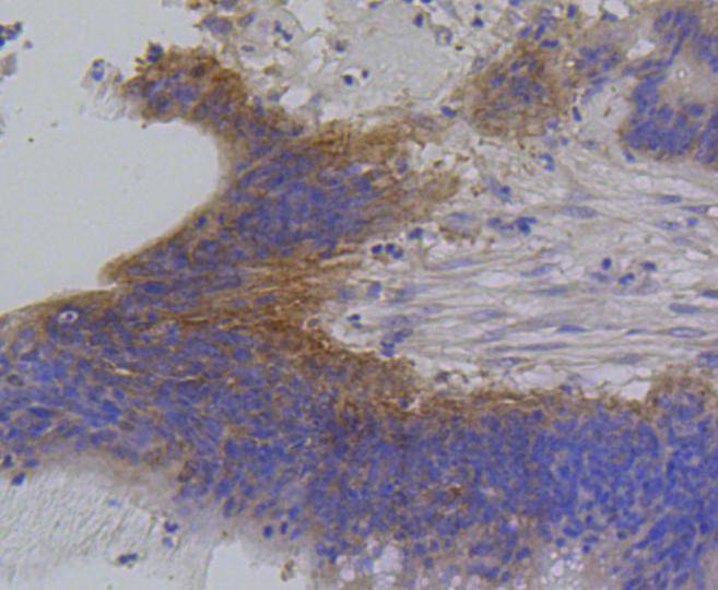
Immunohistochemical analysis of paraffin-embedded human colon cancer tissue using anti-IRF1 antibody. Counter stained with hematoxylin.
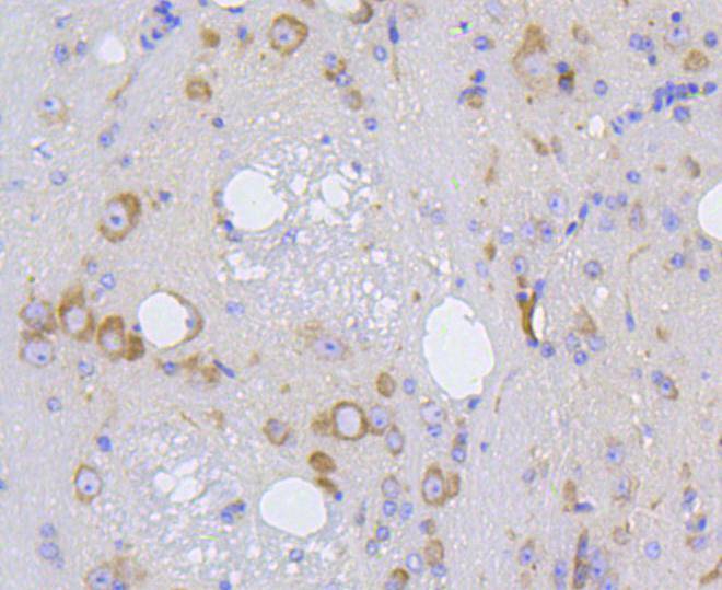
Immunohistochemical analysis of paraffin-embedded mouse brain tissue using anti-IRF1 antibody. Counter stained with hematoxylin.
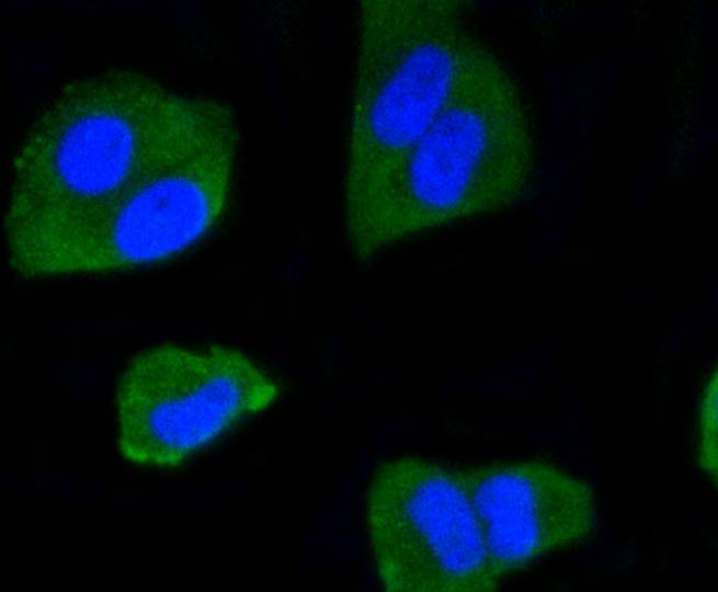
ICC staining IRF1 in Hela cells (green). The nuclear counter stain is DAPI (blue). Cells were fixed in paraformaldehyde, permeabilised with 0.25% Triton X100/PBS.
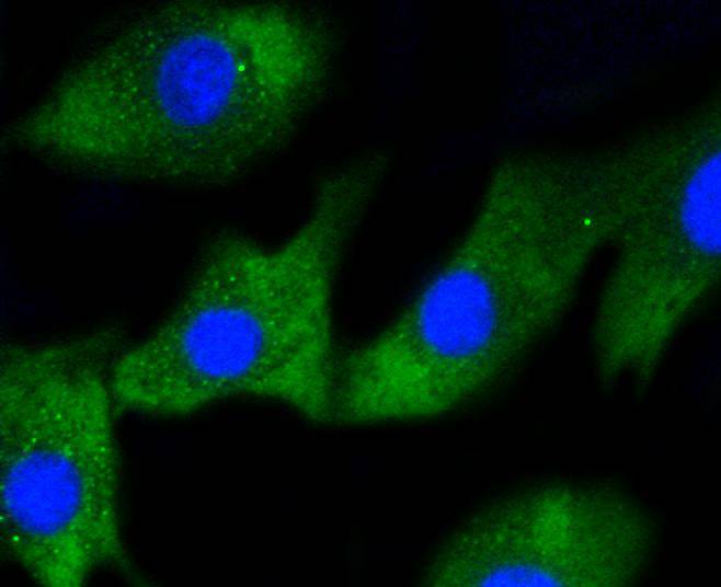
ICC staining IRF1 in NIH/3T3 cells (green). The nuclear counter stain is DAPI (blue). Cells were fixed in paraformaldehyde, permeabilised with 0.25% Triton X100/PBS.
Flow cytometric analysis of Jurkat cells with IRF1 antibody at 1/50 dilution (blue) compared with an unlabelled control (cells without incubation with primary antibody; red). Alexa Fluor 488-conjugated goat anti rabbit IgG was used as the secondary antibody.
背景
Interferon regulatory factor-1 (IRF-1) and IRF-2 have been identified as novel DNA-binding factors that function as regulators of both type I interferon (interferon- and ��) and interferon-inducible genes. The two factors are structurally related, particularly in their N-terminal regions, which confer DNA binding specificity. In addition, both bind to the same sequence within the promoters of interferon- and interferon-�� genes. IRF-1 functions as an activator of interferon transcription, while IRF-2 binds to the same cis elements and represses IRF-1 action. IRF-1 and IRF-2 have been reported to act in a mutually antagonistic manner in regulating cell growth; overexpression of the repressor IRF-2 leads to cell transformation while concomitant overexpression of IRF-1 causes reversion. IRF-1 and IRF-2 are members of a larger family of DNA binding proteins that includes IRF-3, IRF-4, IRF-5, IRF-6, IRF-7, ISGF-3 p48 (a component of the ISGF-3 complex) and IFN consensus sequence-binding protein (ICSBP).
背景文献
1. Maarifi, G. et al. 2015. Small Ubiquitin-like Modifier Alters IFN Response. Journal of immunology (Baltimore, Md. : 1950). 195: 2312-24.
2. Aguado, L.C. et al. 2015. microRNA Function Is Limited to Cytokine Control in the Acute Response to Virus Infection. Cell host & microbe. 18: 714-22.


 Yes
Yes