产品详情
产品名称CDK1 Rabbit mAb
来源种属Recombinant Rabbit
克隆性 Monoclonal antibody
克隆性SM01-44
纯化ProA affinity purified
应用WB, ICC/IF, IHC, IP
种属反应性Hu, Ms, Rt
免疫原描述recombinant protein
别名Cdc 2 antibody Cdc2 antibody CDC28A antibody CDK 1 antibody CDK1 antibody CDK1_HUMAN antibody CDKN1 antibody CELL CYCLE CONTROLLER CDC2 antibody Cell division control protein 2 antibody Cell division control protein 2 homolog antibody Cell division cycle 2 G1 to S and G2 to M antibody Cell division protein kinase 1 antibody Cell Divsion Cycle 2 Protein antibody Cyclin Dependent Kinase 1 antibody Cyclin-dependent kinase 1 antibody DKFZp686L20222 antibody MGC111195 antibody p34 Cdk1 antibody p34 protein kinase antibody P34CDC2 antibody
数据库入口号Swiss-Prot#:P06493
计算分子量34 kDa
配方1*TBS (pH7.4), 1%BSA, 40%Glycerol. Preservative: 0.05% Sodium Azide.
保存Store at -20˚C
应用详情
WB: 1:1,000-1:2,000
IHC: 1:50-1:200
ICC: 1:50-1:200
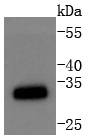
Western blot analysis of CDK1 on Jurkat cells lysates using anti-CDK1 antibody at 1/1,000 dilution.
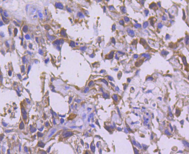
Immunohistochemical analysis of paraffin-embedded human breast carcinoma tissue using anti-CDK1 antibody. Counter stained with hematoxylin.
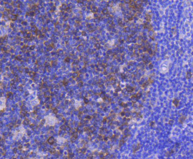
Immunohistochemical analysis of paraffin-embedded human tonsil tissue using anti-CDK1 antibody. Counter stained with hematoxylin.
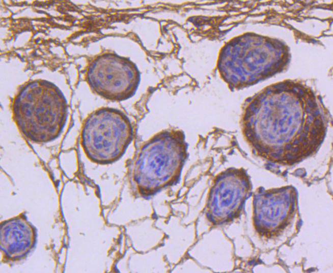
Immunohistochemical analysis of paraffin-embedded mouse skin tissue using anti-CDK1 antibody. Counter stained with hematoxylin.
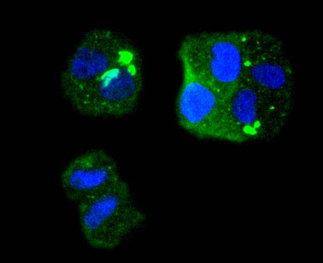
ICC staining CDK1 in Hela cells (green). The nuclear counter stain is DAPI (blue). Cells were fixed in paraformaldehyde, permeabilised with 0.25% Triton X100/PBS.
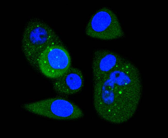
ICC staining CDK1 in MCF-7 cells (green). The nuclear counter stain is DAPI (blue). Cells were fixed in paraformaldehyde, permeabilised with 0.25% Triton X100/PBS.
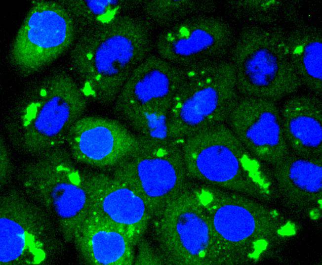
ICC staining CDK1 in A431 cells (green). The nuclear counter stain is DAPI (blue). Cells were fixed in paraformaldehyde, permeabilised with 0.25% Triton X100/PBS.
背景
Cdk1 is a small protein (approximately 34 kilodaltons), and is highly conserved. Cdk1 is comprised mostly by the bare protein kinase motif, which other protein kinases share. Cdk1, like other kinases, contains a cleft in which ATP fits. When bound to its cyclin partners, Cdk1 phosphorylation leads to cell cycle progression. Given its essential role in cell cycle progression, Cdk1 is highly regulated. Most obviously, Cdk1 is regulated by its binding with its cyclin partners. Cyclin binding alters access to the active site of Cdk1, allowing for Cdk1 activity; furthermore, cyclins impart specificity to Cdk1 activity. At least some cyclins contain a hydrophobic patch which may directly interact with substrates, conferring target specificity. Furthermore, cyclins can target Cdk1 to particular subcellular locations.
背景文献
1. Gao K et al. HDGF-related protein-2 (HRP-2) acts as an oncogene to promote cell growth in hepatocellular carcinoma. Biochem Biophys Res Commun 458:849-55 (2015). 2. Wang JF et al. The molecular mechanisms of Tanshinone IIA on the apoptosis and arrest of human esophageal carcinoma cells. Biomed Res Int 2014:582730 (2014).
如果您使用该产品48788发表了文章,请通知我们,让我们可以引用您的文献。
et al,Apoptosis-inducing activity of synthetic hydrocarbon-stapled peptides in H358 cancer cells expressing KRASG12C. In Acta Pharm Sin B on 2021 Sep by Cuicui Li, Ni Zhao,et al..PMID:34589388,
, (2021),
PMID: 34589388
et al,Selective apoptosis-inducing activity of synthetic hydrocarbon-stapled SOS1 helix with d-amino acids in H358 cancer cells expressing KRASG12C.In Eur J Med Chem on 2020 Jan 1 by Xu LL, Li CC et al..PMID:31706640,
, (2020),
PMID: 31706640


 Yes
Yes