免费电话:400-9961-933
邮箱:techcn@signalwayantibody.com
欢迎光临本站
登录 注册 English
位置: 首页 > 重组兔单抗 > Prion Protein(PrP) Rabbit mAb

Prion Protein(PrP) Rabbit mAb#48939

评价
Recombinant
Prion Protein(PrP) Rabbit mAb
是否有货: Yes
产品总价:
产品详情

产品名称Prion Protein(PrP) Rabbit mAb

克隆性SC57-05

纯化ProA affinity purified

应用WB, ICC/IF, IHC, FC

种属反应性Hu, Ms, Rt

免疫原描述recombinant protein

别名Alternative prion protein; major prion protein antibody
AltPrP antibody
ASCR antibody
CD230 antibody
CD230 antigen antibody
CJD antibody
GSS antibody
KURU antibody
Major prion protein antibody
p27 30 antibody
PRIO_HUMAN antibody
Prion protein antibody
Prion related protein antibody
PRIP antibody
PRNP antibody
PrP antibody
PrP27 30 antibody
PrP27-30 antibody
PrP33-35C antibody
PrPC antibody
PrPSc antibody
Sinc antibody

数据库入口号Swiss-Prot#:P04156

计算分子量28 kDa

浓度0.4 mg/ml

配方1*TBS (pH7.4), 1%BSA, 40%Glycerol. Preservative: 0.05% Sodium Azide.

保存Store at -20˚C

应用详情

WB: 1:1,000-5,000
IHC: 1:50-1:200
ICC: 1:50-1:200

FC: 1:50-1:100

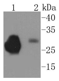
Western blot analysis of PrP on different lysates using anti-PrP antibody at 1/1,000 dilution. Positive control:
Lane 1: Rat brain
Lane 2: Mouse brain
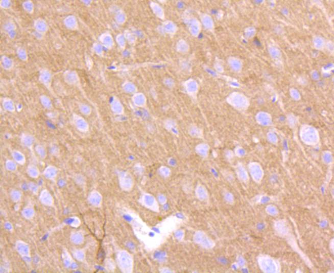
Immunohistochemical analysis of paraffin-embedded rat brain tissue using anti-PrP antibody. Counter stained with hematoxylin.
Immunohistochemical analysis of paraffin-embedded mouse brain tissue using anti-PrP antibody. Counter stained with hematoxylin.
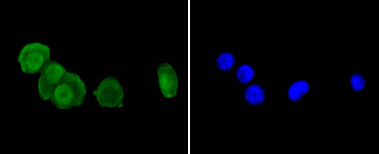
ICC staining PrP in N2A cells (green). The nuclear counter stain is DAPI (blue). Cells were fixed in paraformaldehyde, permeabilised with 0.25% Triton X100/PBS.
ICC staining PrP in SHG-44 cells (green). The nuclear counter stain is DAPI (blue). Cells were fixed in paraformaldehyde, permeabilised with 0.25% Triton X100/PBS.
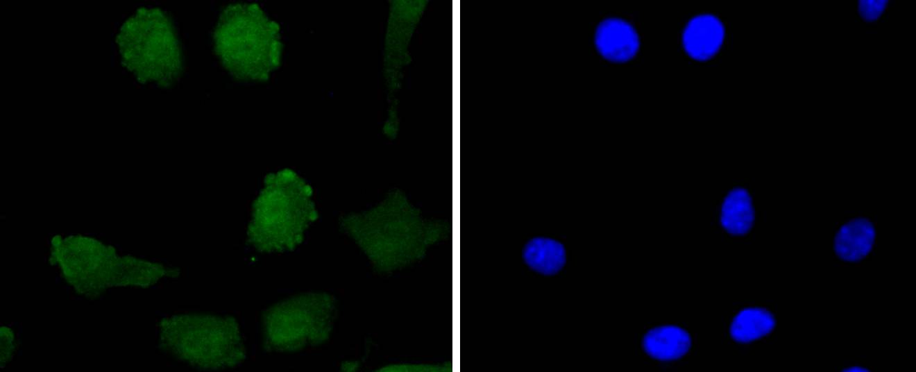
ICC staining PrP in SH-SY-5Y cells (green). The nuclear counter stain is DAPI (blue). Cells were fixed in paraformaldehyde, permeabilised with 0.25% Triton X100/PBS.
Flow cytometric analysis of SH-SY-5Y cells with PrP antibody at 1/50 dilution (red) compared with an unlabelled control (cells without incubation with primary antibody; black). Alexa Fluor 488-conjugated goat anti rabbit IgG was used as the secondary antibody.
背景

Prion diseases, or transmissible spongiform encephalopathies (TSEs), are manifested as genetic, infectious or sporadic, lethal neurodegenerative disorders involving alterations of the prion protein (PrP). Characteristic of prion diseases, cellular PrP (PrPc) is converted to the disease form, PrPSc, through alterations in the protein folding conformations. PrPc is constitutively expressed in normal adult brain and is sensitive to proteinase K digestion, while the altered PrPSc conformation is resistant to proteases, resulting in a distinct molecular mass after PK treatment. Consistent with the transient infection process of prion diseases, incubation of PrPc with PrPSc both in vitro and in vivo produces PrPc that is resistant to protease degradation. Infectious PrPSc is found at high levels in the brains of animals affected by TSEs, including scrapie in sheep, BSE in cattle and Cruetzfeldt-Jakob disease in humans.

背景文献

1. Rouvinski A et al. Live imaging of prions reveals nascent PrPSc in cell-surface, raft-associated amyloid strings and webs. J Cell Biol 204:423-41 (2014).
2. Liu T et al. IGF-1-induced enhancement of PRNP expression depends on the negative regulation of transcription factor FOXO3a. PLoS One 8:e71896 (2013).

如果您使用该产品48939发表了文章,请通知我们,让我们可以引用您的文献。

5

注释

应用

  • WB免疫印迹
  • IHC免疫组化
  • IF免疫荧光
  • ICC免疫细胞化学
  • FC流式细胞
  • IP免疫沉淀
  • E酶联免疫吸附法
  • DB免疫斑点法
  • ChIP染色质免疫沉淀
  • GICA胶体金免疫层析法
  • NC阴性对照

种属反应性

  • Hu
  • Ms小鼠
  • Rt大鼠
  • Dm果蝇
  • C线虫
  • Mk
  • Rb
  • B
  • D
  • P
  • Hm仓鼠
  • ChHm中国仓鼠
  • Chk

联系我们

Location :

南京市高新开发区星火路10号人才大厦C座

Phone :

400-9961-933
025-58868422

Email Address

techcn@signalwayantibody.com