产品详情
产品名称NUR77 Rabbit mAb
克隆性ET1703-97
纯化ProA affinity purified
应用WB, ICC/IF, IHC
种属反应性Hu, Ms, Rt
免疫原描述Synthetic peptide within Human NUR77 aa 10-49 / 598.
别名Early response protein NAK1 antibody
GFRP 1 antibody
GFRP antibody
GFRP1 antibody
Growth factor inducible nuclear protein N10 antibody
Growth Factor Inducible Nuclear Protein NP10 antibody
Growth Factor Response Protein 1 antibody
Hbr1 antibody
HMR antibody
Hormone Receptor antibody
MGC9485 antibody
N10 antibody
N10 nuclear protein antibody
NAK 1 antibody
NAK1 antibody
Nerve growth factor IB nuclear receptor variant 1 antibody
NGFIB antibody
NP 10 antibody
NP10 antibody
NR4A1 antibody
NR4A1_HUMAN antibody
Nuclear hormone receptor NUR/77 antibody
Nuclear Hormone Receptor TR3 antibody
Nuclear receptor subfamily 4 group A member 1 antibody
NUR77 antibody
NUR77, mouse, homolog of antibody
Orphan nuclear receptor HMR antibody
Orphan nuclear receptor NR4A1 antibody
Orphan nuclear receptor TR3 antibody
Orphan receptor tr3 antibody
Receptor NGFIB antibody
ST 59 antibody
ST-59 antibody
ST59 antibody
Steroid receptor TR3 antibody
Testicular receptor 3 antibody
TR 3 antibody
TR3 antibody
TR3 orphan receptor antibody
数据库入口号Swiss-Prot#:P22736
计算分子量64 kDa
配方1*TBS (pH7.4), 0.05% BSA, 40% Glycerol. Preservative: 0.05% Sodium Azide.
保存Store at -20˚C
应用详情
WB: 1:500-1:1000
IHC: 1:50-1:200
ICC: 1:50-1:200
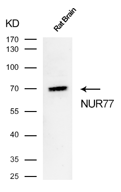
Western blot analysis of NUR77 on rat brain cells lysates using anti-NUR77 antibody at 1/500 dilution.
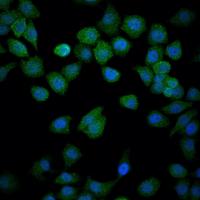
ICC staining of NUR77 in Hela cells (green).4% PFA fixed cells 20 minutes, washed with PBS. Cells were probed with the primary antibody (49510,1/50) overnight at 4℃, washed with PBS.CoraLite488 Goat anti-Rabbit lgG was used as the secondary antibody at 1/100 dilution.The nuclear counter stain is DAPI (blue).
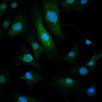
ICC staining of NUR77 in 3T3-L1 cells (green).4% PFA fixed cells 20 minutes, washed with PBS. Cells were probed with the primary antibody (49510,1/50) overnight at 4℃, washed with PBS.CoraLite488 Goat anti-Rabbit lgG was used as the secondary antibody at 1/100 dilution.The nuclear counter stain is DAPI (blue).
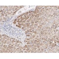
Immunohistochemical analysis of paraffin-embedded human liver tissue with Rabbit anti-NUR77 antibody (ET1703-97) at 1/1,000 dilution.
The section was pre-treated using heat mediated antigen retrieval with sodium citrate buffer (pH 6.0) for 2 minutes. The tissues were blocked in 1% BSA for 20 minutes at room temperature, washed with ddH2O and PBS, and then probed with the primary antibody at 1/1,000 dilution for 1 hour at room temperature. The detection was performed using an HRP conjugated compact polymer system. DAB was used as the chromogen. Tissues were counterstained with hematoxylin and mounted with DPX.
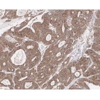
Immunohistochemical analysis of paraffin-embedded human colon carcinoma tissue with Rabbit anti-NUR77 antibody at 1/1,000 dilution.
The section was pre-treated using heat mediated antigen retrieval with sodium citrate buffer (pH 6.0) for 2 minutes. The tissues were blocked in 1% BSA for 20 minutes at room temperature, washed with ddH2O and PBS, and then probed with the primary antibody at 1/1,000 dilution for 1 hour at room temperature. The detection was performed using an HRP conjugated compact polymer system. DAB was used as the chromogen. Tissues were counterstained with hematoxylin and mounted with DPX.
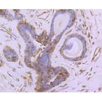
Immunohistochemical analysis of paraffin-embedded human breast tissue using anti-NUR77 antibody. The section was pre-treated using heat mediated antigen retrieval with Tris-EDTA buffer (pH 8.0-8.4) for 20 minutes.The tissues were blocked in 5% BSA for 30 minutes at room temperature, washed with ddH2O and PBS, and then probed with the primary antibody (1/50) for 30 minutes at room temperature. The detection was performed using an HRP conjugated compact polymer system. DAB was used as the chromogen. Tissues were counterstained with hematoxylin and mounted with DPX.
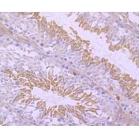
Immunohistochemical analysis of paraffin-embedded human breast tissue using anti-NUR77 antibody. The section was pre-treated using heat mediated antigen retrieval with Tris-EDTA buffer (pH 8.0-8.4) for 20 minutes.The tissues were blocked in 5% BSA for 30 minutes at room temperature, washed with ddH2O and PBS, and then probed with the primary antibody (1/50) for 30 minutes at room temperature. The detection was performed using an HRP conjugated compact polymer system. DAB was used as the chromogen. Tissues were counterstained with hematoxylin and mounted with DPX.
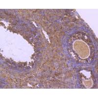
Immunohistochemical analysis of paraffin-embedded mouse ovarian tissue using anti-NUR77 antibody. The section was pre-treated using heat mediated antigen retrieval with Tris-EDTA buffer (pH 8.0-8.4) for 20 minutes.The tissues were blocked in 5% BSA for 30 minutes at room temperature, washed with ddH2O and PBS, and then probed with the primary antibody (1/50) for 30 minutes at room temperature. The detection was performed using an HRP conjugated compact polymer system. DAB was used as the chromogen. Tissues were counterstained with hematoxylin and mounted with DPX.
背景
Nurr1 (Nur-related factor 1) and Nur77 (also designated NGFI-B) encode orphan nuclear receptors which may comprise an additional subfamily within the nuclear receptor superfamily. The rat and human homologs of mouse Nurr1 are designated RNR1 and NOT, respectively. Both Nurr1 and Nur77 are growth factor inducible immediate early response genes. Induction of both Nurr1 and Nur77 is seen after membrane depolarization while only Nur77 induction is seen with NGF stimulation. JunD acts as a mediator for Nur77. An increase in Nur77 expression is seen in activated T cells during G0 to G1 transition and throughout the G1 phase. In addition to its function as an immediate early gene, Nur77 may play a role in TCR-mediated apoptosis. Cyclosporin A, a potent immunosuppressant, has been shown to inhibit the ability of Nur77 to bind DNA. A dominant negative form of Nur77 can protect T cell hybridomas from activation-induced apoptosis. However, the absolute requirement of Nur77 for TCR-mediated apoptosis is still under debate.
1. Liu NA et al. Cyclin E - mediated human proopiomelanocortin regulation as a therapeutic target for Cushing disease. J Clin Endocrinol Metab N/A:jc20151606 (2015).
2. Yu Y et al. The orphan nuclear receptor Nur77 inhibits low shear stress-induced carotid artery remodeling in mice. Int J Mol Med 36:1547-55 (2015).


 Yes
Yes

