产品详情
产品名称CD146 Rabbit mAb
克隆性JB42-35
纯化ProA affinity purified
应用WB,ICC,IF,IHC,FC
种属反应性Hu, Ms, Rt
免疫原描述Recombinant protein
别名A32 antigen antibody
CD 146 antibody
CD146 antibody
CD146 antigen antibody
Cell surface glycoprotein MUC18 antibody
Cell surface glycoprotein P1H12 antibody
Gicerin antibody
Mcam antibody
Melanoma adhesion molecule antibody
Melanoma associated antigen A32 antibody
Melanoma associated antigen MUC18 antibody
Melanoma associated glycoprotein MUC18 antibody
Melanoma cell adhesion molecule antibody
Melanoma-associated antigen A32 antibody
Melanoma-associated antigen MUC18 antibody
MelCAM antibody
MUC 18 antibody
MUC18 antibody
MUC18_HUMAN antibody
S endo 1 antibody
S endo 1 endothelial associated antigen antibody
S-endo 1 endothelial-associated antigen antibody
数据库入口号Swiss-Prot#:P43121
计算分子量72 kDa
配方1*TBS (pH7.4), 1%BSA, 40%Glycerol. Preservative: 0.05% Sodium Azide.
保存Store at -20˚C
应用详情
WB: 1:500-1:1,000
IHC: 1:50-1:200
ICC: 1:100-1:500
FC: 1:50-1:100
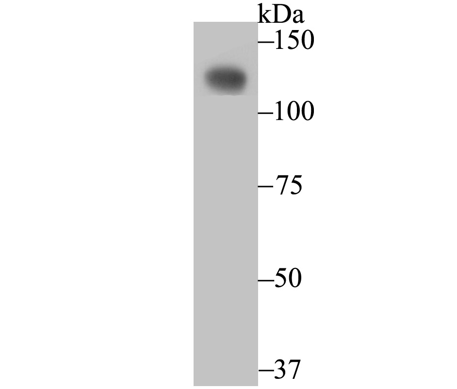
Western blot analysis of CD146 on SiHa cell using anti-CD146 antibody at 1/500 dilution.
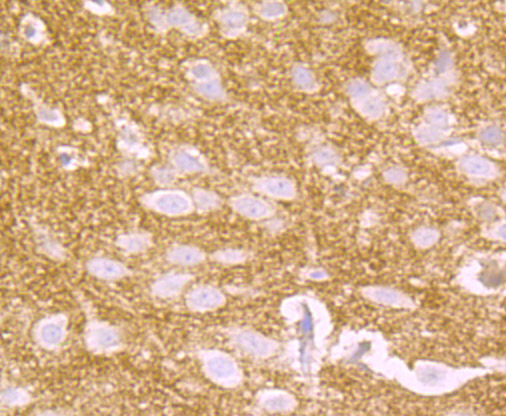
Immunohistochemical analysis of paraffin-embedded rat brain tissue using anti-CD146 antibody. Counter stained with hematoxylin.
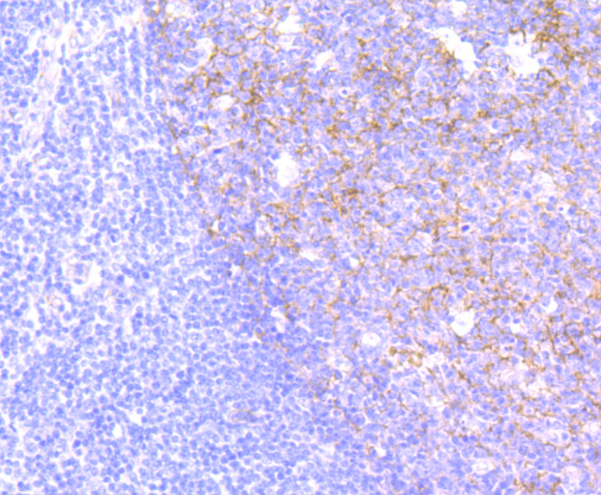
Immunohistochemical analysis of paraffin-embedded human tonsil tissue using anti-CD146 antibody. Counter stained with hematoxylin.
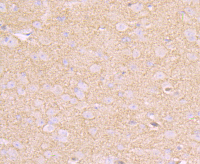
Immunohistochemical analysis of paraffin-embedded mouse brain tissue using anti-CD146 antibody. Counter stained with hematoxylin.
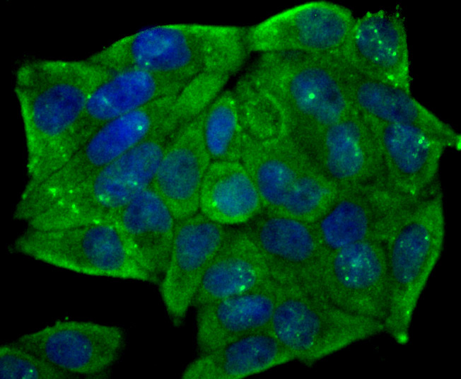
ICC staining CD146 in Hela cells (green). The nuclear counter stain is DAPI (blue). Cells were fixed in paraformaldehyde, permeabilised with 0.25% Triton X100/PBS.
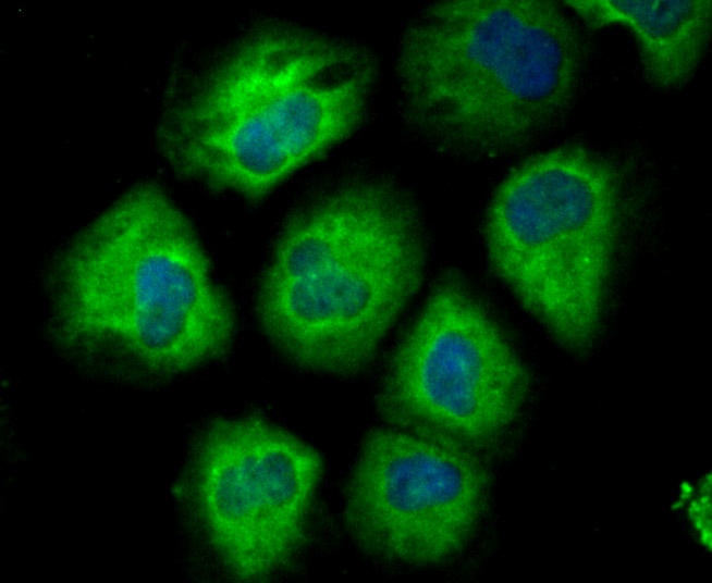
ICC staining CD146 in HUVEC cells (green). The nuclear counter stain is DAPI (blue). Cells were fixed in paraformaldehyde, permeabilised with 0.25% Triton X100/PBS.
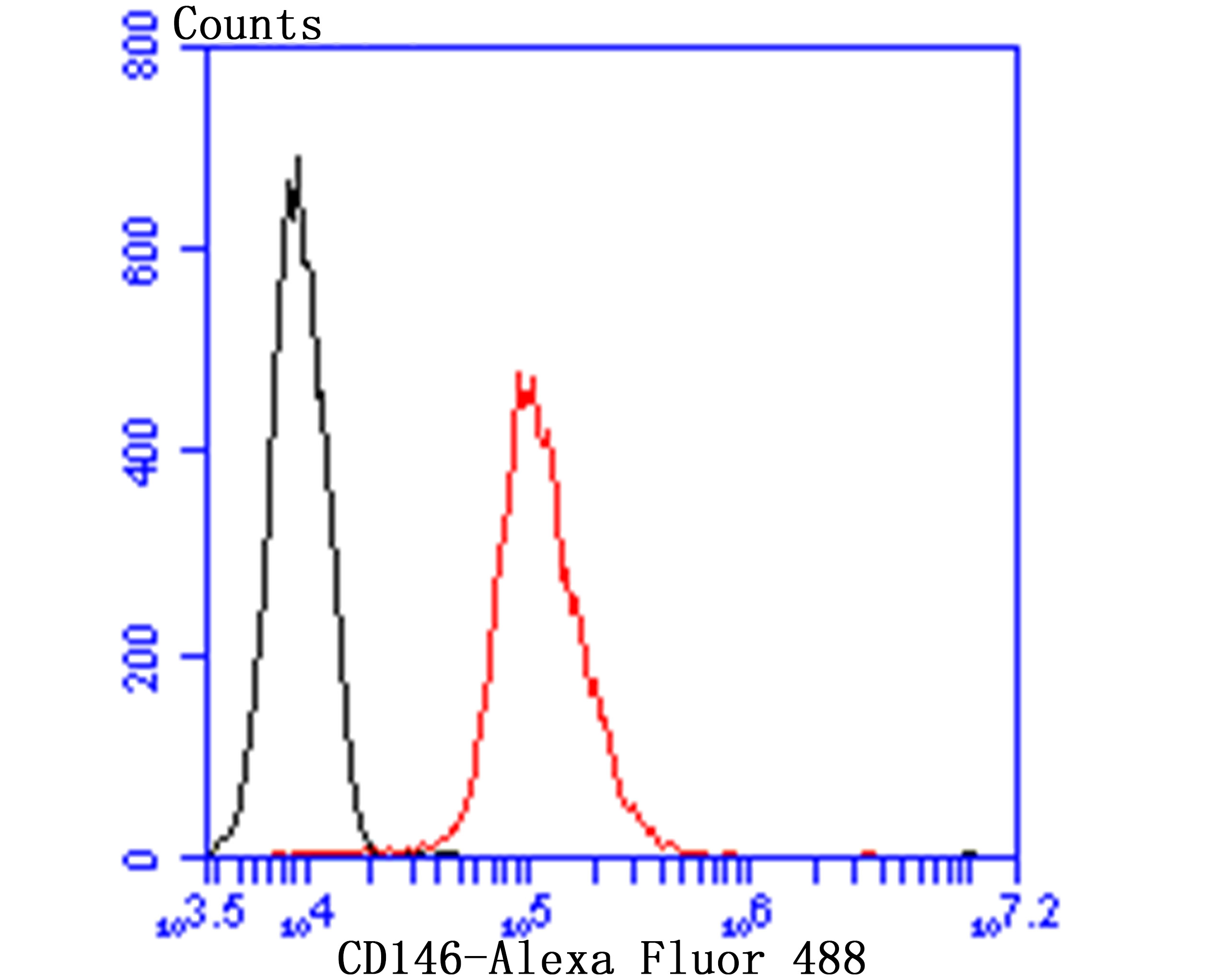
Flow cytometric analysis of HUVEC cells with CD146 antibody at 1/100 dilution (red) compared with an unlabelled control (cells without incubation with primary antibody; black). Alexa Fluor 488-conjugated goat anti-rabbit IgG was used as the secondary antibody.
背景
The tumorigenic and metastatic phenotype of melanoma cells correlates well with an increased expression of cell-cell and cell-matrix adhesion receptors. The human Mel-CAM gene encodes a transmembrane glycoprotein, also designated MCAM, MUC18 or CD146, that belongs to the immunoglobulin superfamily and functions as a Ca2+-independent cell adhesion molecule. The deduced human sequence of 603 amino acids consists of a signal peptide, five immunoglobulin-like domains, a transmembrane region and a short cytoplasmic tail. Mel-CAM expression is restricted to advanced primary and metastatic melanomas and to cell lines of the neuroectodermal lineage, but not normal melanocytes. Mel-CAM is found on 80% of advanced primary human mela-nomas and correlates well with development of metastatic disease. Mel-CAM activation initiates an outside-in signaling pathway that involves the protein tyrosine kinases Fyn, FAK and paxillin. Mel-CAM influences the dynamics of Actin cytoskeleton rearrangement and is essential for the maintenance of thymic architecture and function.
1. Johnson J P et al. The progression associated antigen MUC18: a unique member of the immunoglobulin supergene family. Melanoma Res 3:337-340 (1993).
2. Anfosso F et al. Outside-in signaling pathway linked to CD146 engagement in human endothelial cells. J Biol Chem 276:1564-1569 (2001).


 Yes
Yes