The biological activities of IL-2 are mediated by its binding to a multi-molecular cellular receptor complex. For several years the receptor was thought to consist of two glycoprotein chains, an alpha chain (IL-2 Rα) and a beta chain (IL-2 Rβ) (1 - 3), which acted together to form a high affinity receptor that transduced the IL-2 signal. IL-2 Rα (also known as Tac antigen and as CD25) is a 55 kDa transmembrane glycoprotein composed of 351 amino acids with only 13 located on the cytoplasmic side of the membrane (4 - 6). The second chain of the complex was cloned in 1989 (7), and is a transmembrane glycoprotein of 575 amino acids (75 kDa), 286 of which are located cytoplasmically and clearly participate in signal transduction (8, 9). Eventually it was discovered that a third chain, IL-2 Rγ,
was necessary for high affinity binding, ligand internalization and signalling. Constitutively expressed on many lymphoid cells, it had been overlooked partly because it has no affinity for IL-2 except when IL-2 Rβ is present (7, 10, 11). When cloned, the gene was found to code for a 64 kDa transmembrane protein of 347 amino acids, 84 of which are cytoplasmic (12). Both IL-2 Rβ and IL-2 Rγ are members of the hematopoietin receptor superfamily, whereas IL-2 Rα is related only to the IL-15 R αchain (13 - 15).
A model of the IL-2 receptor complex (3, 9, 16 - 21) would describe the high affinity receptor as an αβγ trimer, in which all three chains are in contact with the ligand. Alone, IL-2 Rα binds IL-2 with low affinity, but is unable to transduce a signal. The αβ combination will bind IL-2 with intermediate affinity, but still will not transduce a signal. A βγ complex has intermediate affinity and is capable of signalling if the IL-2 concentration is relatively high. Regardless of many subtleties that determine the affinity of the ligand for the extracellular portions of the receptor components (22 - 24), signalling will ensue if the βand γcytoplasmic domains are brought into close proximity (25 - 27).
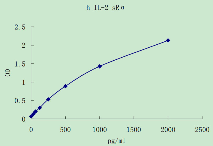
A soluble form of IL-2 Rα appears in serum, concomitant with its increased expression on cells (18, 28, 29). There are reports of a soluble form of IL-2 Rβ as well (28, 30). The function of the soluble IL-2 Rα is unclear, since it would be expected to be a poor inhibitor of IL-2 because of its low binding affinity. In any case, increased levels of the soluble IL-2 Rα in biological fluids reportedly correlate with increased T and B cell activation and immune system activation. Results of a number of studies suggest a correlation of levels of IL-2 sRα in serum with the onset of rejection episodes in allograft recipients (18, 31 - 33), with activity of autoimmune diseases such as rheumatoid arthritis and systemic lupus erythematosis (SLE) (34) and with the course of some leukemias and lymphomas (35 - 40).
Tsudo, M. et al. (1986) Proc. Natl. Acad. Sci. USA 83:9694.
Sharon, M. et al. (1986) Science 234:859.
Smith, K.A. (1989) Annu. Rev. Cell Biol. 5:397.
Leonard, W.J. et al. (1984) Nature 311:626.
Nikaido, T. et al. (1984) Nature 311:631.
Cosman, D. et al. (1984) Nature 312:768.
Hatakeyama, M. et al. (1989) Science 244:551.
Hatakeyama, M. et al. (1991) Science 252:1523.
Minami, Y. et al. (1993) Annu. Rev. Immunol. 11:245.
Hatakeyama, M. et al. (1985) Nature 318:467.
Zurawsky, S.M. et al. (1990) EMBO J. 9:3899.
Takeshita, T. et al. (1992) Science 257:379.
Cosman, D. et al. (1993) Cytokine 5:95.
Bazan, J.F. (1990) Proc. Natl. Acad. Sci. USA 87:6934.
Anderson, D.M. (1995) J. Biol. Chem. 270:29862.
Taniguchi, T. and Y. Minami (1993) Cell 73:5.
Waldmann, T.A. (1991) J. Biol. Chem. 266:2681.
Waldmann, T.A. (1993) Immunol. Today 14:264.
Voss, S.D. et al. (1994) Blood 83:626.
Leonard, W.J. et al. (1994) Immunol. Rev. 138:61.
Leonard, W.J. et al. (1994) Curr. Opin. Immunol. 6:631.
Voss, S.D. et al. (1993) Proc. Natl. Acad. Sci. USA 90:2428.


 1-2周
1-2周