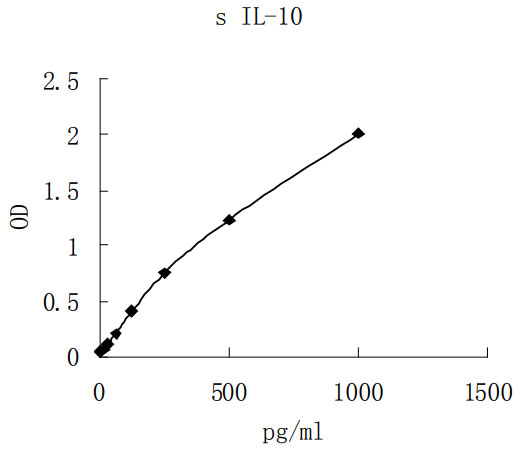
产品名称Swine IL-10 ELISA kit
简述ELISA Kit
应用ELISA
种属反应性Swine
特异性Natural and recombinant Swine IL-10 Ligand
交叉反应性No significant interference observed with available related molecules.
基因/蛋白名称Swine IL-10
Sensitivity: 7pg/mL
Sample Type: Cell culture supernatant, serum, plasma (EDTA, citrate, heparin)
Sample Volume: 20 uL
Assay Time: 3 hour
Detection method: Colorimetric

- Aluminium pouches with a Microwell Plate coated with antibody to swine IL-10 (8X12)
- 2 vials swine IL-10 Standard lyophilized, 1000 pg/ml upon reconstitution
- 2 vials concentrated Biotin-Conjugate anti-swine IL-10 antibody
- 2 vials Streptavidin-HRP solution,
- 1 bottle Standard /sample Diluent
- 1 bottle Biotin-Conjugate antibody Diluent
- 1 bottle Streptavidin-HRP Diluent
- 1 bottle Wash Buffer Concentrate 20x (PBS with 1% Tween-20)
- 1 vial Substrate Solution
- 1 vial Stop Solution
- 4 pieces Adhesive Films
- package insert
Interleukin 10, also known as cytokine synthesis inhibitory factor (CSIF), is the charter member of the IL-10 cytokine family. This family currently comprises IL-10, IL-19, IL-20, IL-22, IL-24, and IL-26/AK155 (1-4). All IL-10 family members are secreted ?-helical proteins. Porcine IL-10 is a secreted, possibly glycosylated, polypeptide with an 18 kDa molecular weight (5). Based on human studies, porcine IL-10 is likely to circulate as a nondisulfide-linked homodimer (3). Porcine IL-10 is synthesized as a 175 amino acid (aa) precursor with an 18 aa signal sequence and a 157 aa mature form. The mature segment has one potential N-linked glycosylation site plus four cysteines which form two intrachain disulfide bridges (6). Mature porcine IL-10 shows 71%, 70%, 76%, 75%, 77%, and 71% aa sequence identity to rat (7), mouse (8), human (9), guinea pig (10), canine (11), and cotton rat (12) IL-10, respectively. Upon activation, mammalian cells known to secrete IL-10 include NK cells (13), cytotoxic CD8+ T cells secreting Th2-like cytokines (14), CD4+CD45RA- (memory) Th1 and Th2 cells (15), macrophages (16), monocytes (17), CD5+ and CD5- B cells (18, 19), dendritic cells (20, 21), hepatic stellate (Ito cells) (22), keratinocytes (23), melanoma cells (24), mast cells (25), placental cytotrophoblasts (26), and fetal erythroblasts (27).
The functional receptor for IL-10 (IL-10 R) in pigs has not been reported. By analogy to human, it would be expected to be composed of two 110 kDa -chains (or IL-10 R1) and two 75 kDa -chains (or IL-10 R2) (28 - 31). The -chain binds IL-10 and transduces a signal in the presence of a -chain complex (29). Both receptors are members of the class II cytokine receptor family (CRF2) that is characterized by the presence of type III fibronectin domains and conserved tryptophans (29, 31). This class does not possess the WSXWS motif characteristic of the class I CRF. There is no significant aa sequence identity (< 30%) between human IL-10 R1 and IL-10 R2.
IL-10 has myriad effects on a variety of cell types. On activated B cells, IL-10 can induce plasma cell formation (32) and the secretion of either IgG (33, 34) or IgA (in the presence of TGF- 1 and/or IL-4) (34, 35). In the presence of IL-2, CD56+ NK cells will respond to IL-10 with increased proliferation plus IFN- ?and TNF- secretion (36). Conversely, on macrophages, IL-10 is known to downregulate IL-1, TNF- , and IL-6 production (37). On dendritic cells, IL-10 has been shown to interfere with antigen-presenting cell function by downmodulating stimulatory and co-stimulatory molecules (38, 39). On monocytes, IL-10 is reported to direct monocyte differentiation into cytotoxic CD16+ macrophages rather than antigen-presenting dendritic cells (40-42).
Rich, B.E. and T.S. Kupper (2001) Curr. Biol. 11:R531.
Gruenberg, B.H. et al. (2001) Genes Immun. 2:329.
Moore, K.W. et al. (2001) Annu. Rev. Immunol. 19:683.
Fickenscher, H. et. al. (2002) Trends Immunol. 23:89.
Blancho, G. et al. (1995) Proc. Natl. Acad. Sci. USA 92:2800.
Windsor, W.T. et al. (1993) Biochemistry 32:8807.
Feng, L. et al. (1993) Biochem. Biophys. Res. Commun. 192:452.
Moore, K.W. et al. (1990) Science 248:1230.
Vieira, P. et al. (1991) Proc. Natl. Acad. Sci. USA 88:1172.
Scarozza, A.M. et al. (1998) Cytokine 10:851.
Lu, P. et al. (1995) J. Interf. Cytokine Res. 15:1103.
Langley, R.J. et al. (2001) GenBank Accession #AAK04013.
Mehrotra, P.T. et al. (1998) J. Immunol. 160:2637.
Sad, S. et al. (1995) Immunity 2:271.
Yssel, H. et al. (1992) J. Immunol. 149:2378.
Panuska, J.R. et al. (1995) J. Clin. Invest. 96:2445.
Hodge, S. et al. (1999) Scand. J. Immunol. 49:548.
Spencer, N.F.L. and R.A. Daynes (1997) Int. Immunol. 9:745.
O'Garra, A. et al. (1990) Int. Immunol. 2:821.
Iwasaki, A. and B.L. Kelsall (1999) J. Exp. Med. 190:229.
Rea, D. et al. (2000) Blood 95:3162.
Wang, S.C. et al. (1998) J. Biol. Chem. 273:302.
Grewe, M. et al. (1995) J. Invest. Dermatol. 104:3.
Sato, T. et al. (1996) Clin. Cancer Res. 2:1383.
Ishizuka, T. et al. (1999) Clin. Exp. Allergy 29:1424.
Roth, I. et al. (1996) J. Exp. Med. 184:539.
Sennikov, S.V. et al. (2002) Cytokine 17:221.
Kotenko, S.V. et al. (1997) EMBO J. 16:5894.
Lutfalla, G. et al. (1993) Genomics 16:366.
Gibbs, V.C. and D. Pennica (1997) Gene 186:97.
Liu, Y. et al. (1994) J. Immunol. 152:1821.
Rousset, F. et al. (1995) Int. Immunol. 7:1243.
Briere, F. et al. (1994) J. Exp. Med. 179:757.
Defrance, T. et al. (1992) J. Exp. Med. 175:671.
Marconi, M. et al. (1998) Clin. Exp. Immunol. 112:528.
Carson, W.E. et al. (1995) Blood 85:3577.
Fiorentino, D.F. et al. (1991) J. Immunol. 147:3815.
Qzawa, H. et al. (1996) Eur. J. Immunol. 26:648.
Buelens, C. et al. (1995) Eur. J. Immunol. 25:2668.
Olikowsky, T. et al. (1997) Immunology 91:104.
Calzada-Wack, J.C. et al. (1996) J. Inflamm. 46:78.
Allavena, P. et al. (1998) Eur. J. Immunol. 28:359.
如果您使用该产品EK0529发表了文章,请通知我们,让我们可以引用您的文献。


 1-2周
1-2周

