免费电话:400-9961-933
邮箱:techcn@signalwayantibody.com
欢迎光临本站
登录 注册 English
位置: 首页 > 磷酸化抗体 > c-Cbl(phospho-Tyr700) Antibody

c-Cbl(phospho-Tyr700) Antibody#11549

c-Cbl(phospho-Tyr700) Antibody
是否有货: 有货
产品总价:
产品详情

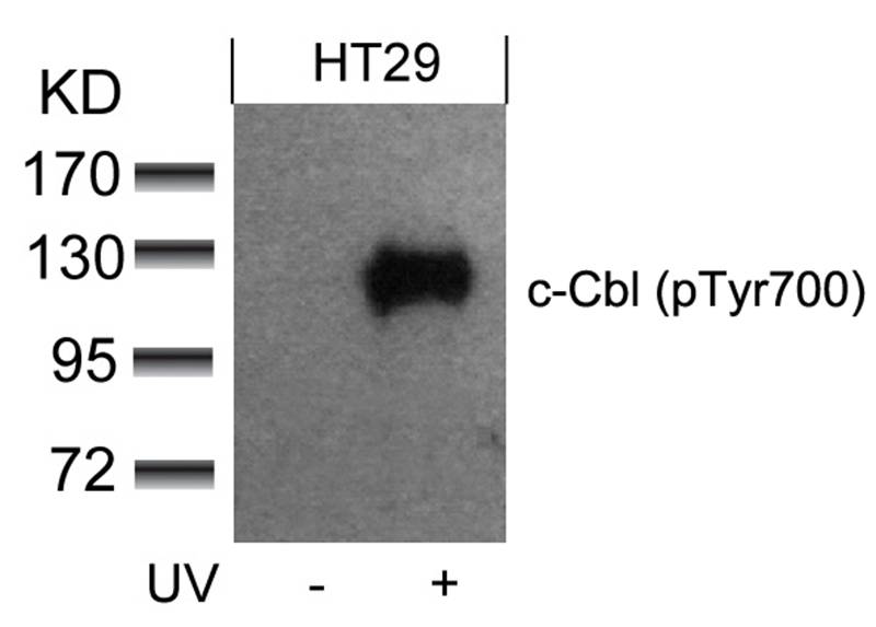
产品名称c-Cbl(phospho-Tyr700) Antibody

来源种属Rabbit

克隆性Polyclonal

纯化Antibodies were produced by immunizing rabbits with synthetic phosphopeptide and KLH conjugates. Antibodies were purified by affinity-chromatography using epitope-specific phosphopeptide. Non-phospho specific antibodies were removed by chromatogramphy using non-phosphopeptide.
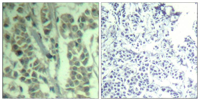
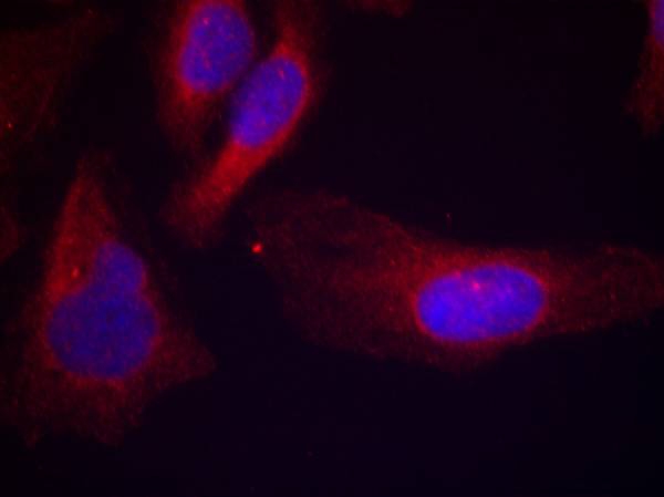
应用WB IHC IF

种属反应性Hu

特异性The antibody detects endogenous level of c-Cbl only when phosphorylated at tyrosine 770.

免疫原类型Peptide-KLH

免疫原描述Peptide sequence around phosphorylation site of tyrosine 770 (T-E-Y(p)-M-T) derived from Human c-Cbl.

基因/蛋白名称c-Cbl

修饰Phospho

别名Signal transduction protein CBL; Proto-oncogene c-CBL; Casitas B-lineage lymphoma proto-oncogene; RING finger protein 55;

数据库入口号Swiss-Prot: P22681
NCBI Protein: NP_005179.2

浓度1.0mg/ml

配方Supplied at 1.0mg/mL in phosphate buffered saline (without Mg2+ and Ca2+), pH 7.4, 150mM NaCl, 0.02% sodium azide and 50% glycerol.

保存Store at -20°C for long term preservation (recommended). Store at 4°C for short term use.

应用详情
Predicted MW: 120kd
Western blotting: 1:500~1:1000
Immunohistochemistry: 1:50~1:100
Immunofluorescence: 1:100~1:200
Western blot analysis of extracts from HT29 cells untreated or treated with UV using c-Cbl(phospho-Tyr700) Antibody #11549.
Immunohistochemical analysis of paraffin-embedded human breast carcinoma tissue using c-Cbl(Phospho-Tyr700) Antibody #11549(left) or the same antibody preincubated with blocking peptide(right).
Immunofluorescence staining of methanol-fixed Hela cells using c-Cbl(phospho-Tyr700) Antibody #11549.
背景
Participates in signal transduction in hematopoietic cells. Adapter protein that functions as a negative regulator of many signaling pathways that start from receptors at the cell surface. Acts as an E3 ubiquitin-protein ligase, which accepts ubiquitin from specific E2 ubiquitin-conjugating enzymes, and then transfers it to substrates promoting their degradation by the proteasome. Recognizes activated receptor tyrosine kinases, including PDGFA, EGF and CSF1, and terminates signaling.

Blake, T.J. et al. (1991) Oncogene 6, 653-657.
Thien, C.B. and Langdon, W.Y. (1998) Immunol. Cell Biol. 76, 473-482.
Kamei, T. et al. (2000) Int. J. Oncol. 17, 335-339.
Hunter, C. et al. (1999) J. Biol. Chem. 274, 2097-2106.

如果您使用该产品11549发表了文章,请通知我们,让我们可以引用您的文献。

5

注释

应用

  • WB免疫印迹
  • IHC免疫组化
  • IF免疫荧光
  • ICC免疫细胞化学
  • FC流式细胞
  • IP免疫沉淀
  • E酶联免疫吸附法
  • DB免疫斑点法
  • ChIP染色质免疫沉淀
  • GICA胶体金免疫层析法
  • NC阴性对照

种属反应性

  • Hu
  • Ms小鼠
  • Rt大鼠
  • Dm果蝇
  • C线虫
  • Mk
  • Rb
  • B
  • D
  • P
  • Hm仓鼠
  • ChHm中国仓鼠
  • Chk

联系我们

Location :

南京市高新开发区星火路10号人才大厦C座

Phone :

400-9961-933
025-58868422

Email Address

techcn@signalwayantibody.com