免费电话:400-9961-933
邮箱:techcn@signalwayantibody.com
欢迎光临本站
登录 注册 English
位置: 首页 > KO/KD抗体 > p53(Ab-15) Antibody

p53(Ab-15) Antibody#21085

评价
KOKD
p53(Ab-15) Antibody
p53(Ab-15) Antibody
p53(Ab-15) Antibody
p53(Ab-15) Antibody
p53(Ab-15) Antibody
p53(Ab-15) Antibody
p53(Ab-15) Antibody
p53(Ab-15) Antibody
p53(Ab-15) Antibody
p53(Ab-15) Antibody
是否有货: 有货
产品总价:
产品详情

产品名称p53(Ab-15) Antibody

来源种属Rabbit

克隆性Polyclonal

纯化Antibodies were produced by immunizing rabbits with synthetic peptide and KLH conjugates. Antibodies were purified by affinity-chromatography using epitope-specific peptide.

应用WB,IHC,IF,ELISA

种属反应性Human,Mouse,Rat,Monkey

特异性The antibody detects endogenous level of total p53 protein.

免疫原类型Peptide-KLH

免疫原描述Peptide sequence around aa.13~17 (P-L-S-Q-E) derived from Human p53.

基因/蛋白名称p53

别名Tumor suppressor p53; Phosphoprotein p53; Antigen NY-CO-13; TP53;

数据库入口号Swiss-Prot: P04637
NCBI Protein: NP_000537.3

浓度1.0mg/ml

配方Supplied at 1.0mg/mL in phosphate buffered saline (without Mg2+ and Ca2+), pH 7.4, 150mM NaCl, 0.02% sodium azide and 50% glycerol.

保存Store at -20°C for long term preservation (recommended). Store at 4°C for short term use.

应用详情

Western Blot: 1:500 - 1:2000.
Immunohistochemistry: 1:100 - 1:300. 
Immunofluorescence: 1:200 - 1:1000

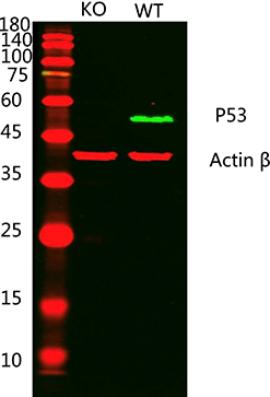
Western blot analysis of lysates from 1)p53 knockout A431 cell , 2)A431 cells, (Green) primary antibody was diluted at 1:1000, 4°over night, Dylight 800 secondary antibody was diluted at 1:10000, 37° 1hour. (Red) Actin β Monoclonal Antibody(5B7) antibody was diluted at 1:5000 as loading control, 4° over night, Dylight 680 secondary antibody was diluted at 1:10000, 37° 1hour.
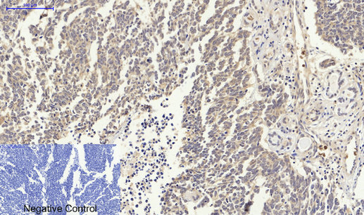
Immunohistochemical analysis of paraffin-embedded Human-lung-cancer tissue. 1,p53 Polyclonal Antibody was diluted at 1:200(4°C,overnight). 2, Sodium citrate pH 6.0 was used for antibody retrieval(>98°C,20min). 3,Secondary antibody was diluted at 1:200(room tempeRature, 30min). Negative control was used by secondary antibody only.
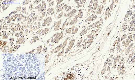
Immunohistochemical analysis of paraffin-embedded Human-stomach-cancer tissue. 1,p53 Polyclonal Antibody was diluted at 1:200(4°C,overnight). 2, Sodium citrate pH 6.0 was used for antibody retrieval(>98°C,20min). 3,Secondary antibody was diluted at 1:200(room tempeRature, 30min). Negative control was used by secondary antibody only.
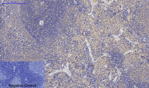
Immunohistochemical analysis of paraffin-embedded Rat-spleen tissue. 1,p53 Polyclonal Antibody was diluted at 1:200(4°C,overnight). 2, Sodium citrate pH 6.0 was used for antibody retrieval(>98°C,20min). 3,Secondary antibody was diluted at 1:200(room tempeRature, 30min). Negative control was used by secondary antibody only.
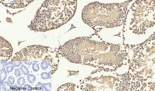
Immunohistochemical analysis of paraffin-embedded Mouse-testis tissue. 1,p53 Polyclonal Antibody was diluted at 1:200(4°C,overnight). 2, Sodium citrate pH 6.0 was used for antibody retrieval(>98°C,20min). 3,Secondary antibody was diluted at 1:200(room tempeRature, 30min). Negative control was used by secondary antibody only.
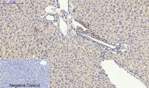
Immunohistochemical analysis of paraffin-embedded Mouse-liver tissue. 1,p53 Polyclonal Antibody was diluted at 1:200(4°C,overnight). 2, Sodium citrate pH 6.0 was used for antibody retrieval(>98°C,20min). 3,Secondary antibody was diluted at 1:200(room tempeRature, 30min). Negative control was used by secondary antibody only.
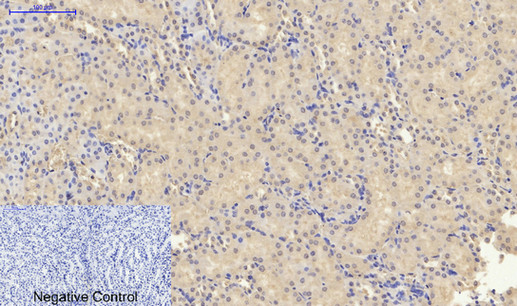
Immunohistochemical analysis of paraffin-embedded Mouse-kidney tissue. 1,p53 Polyclonal Antibody was diluted at 1:200(4°C,overnight). 2, Sodium citrate pH 6.0 was used for antibody retrieval(>98°C,20min). 3,Secondary antibody was diluted at 1:200(room tempeRature, 30min). Negative control was used by secondary antibody only.
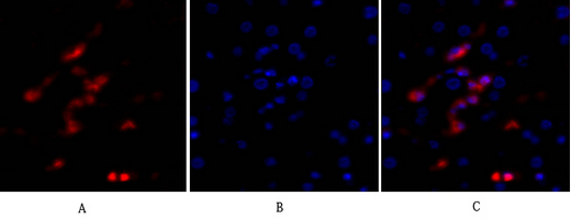
Immunofluorescence analysis of human-liver tissue. 1,p53 Polyclonal Antibody(red) was diluted at 1:200(4°C,overnight). 2, Cy3 labled Secondary antibody was diluted at 1:300(room temperature, 50min).3, Picture B: DAPI(blue) 10min. Picture A:Target. Picture B: DAPI. Picture C: merge of A+B
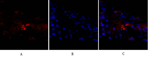
Immunofluorescence analysis of human-lung tissue. 1,p53 Polyclonal Antibody(red) was diluted at 1:200(4°C,overnight). 2, Cy3 labled Secondary antibody was diluted at 1:300(room temperature, 50min).3, Picture B: DAPI(blue) 10min. Picture A:Target. Picture B: DAPI. Picture C: merge of A+B
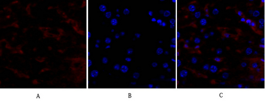
Immunofluorescence analysis of mouse-liver tissue. 1,p53 Polyclonal Antibody(red) was diluted at 1:200(4°C,overnight). 2, Cy3 labled Secondary antibody was diluted at 1:300(room temperature, 50min).3, Picture B: DAPI(blue) 10min. Picture A:Target. Picture B: DAPI. Picture C: merge of A+B
背景

p53 is a nuclear protein which plays an essential role in the regulation of cell cycle specifically in the transition from G0 to G1. It is found in very low levels in normal cells however in a variety of transformed cell lines in high amounts and believed to contribute to transformation and malignancy. The open reading frame of p53 is 393 amino acids long, with the central region (consisting of amino acids from about 100 to 300) containing the DNA-binding domain. This proteolysis-resistant core is flanked by a C-terminal end mediating oligomerization and an N-terminal end containing a strong transcription activation signal. p53 binds as a tetramer to a PBS (p53-Binding Site) and activates the expression of downstream genes that inhibit growth and/or invasion. p53 binds as a tetramer to a p53-binding site (PBS) and to activate the expression of adjacent genes that inhibit growth and/or invasion. Deletion of one or both p53 alleles reduces the expression of tetramers, resulting in decreased expression of the growth inhibitory genes

Lin T, et al. (2005) Nat Cell Biol; 7(2): 165-71.
Vega FM, et al. (2004) Mol Cell Biol; 24(23): 10366-80.
Li J, et al. (2004) J Biol Chem; 279(40): 41275-9.
Wang J, et al. (2004) J Biol Chem; 279(38): 39584-92.

如果您使用该产品21085发表了文章,请通知我们,让我们可以引用您的文献。

et al,CK2-mediated CCDC106 phosphorylation is required for p53 degradation In cancer progression. In J Exp ClIn Cancer Res on 2019 Mar 18 by NIng Y, Wang C, et al..PMID: 30885251, , (2019),
PMID: 30885251
et al,PTEN expression in U251 glioma cells enhances their sensitivity to ionizing radiation by suppressing DNA repair capacity. In Eur Rev Med Pharmacol Sci on 2019 Dec by Li HL, Wang CY, et al..PMID:31841199, , (2019),
PMID: 31841199
et al,Solanesol protects human hepatic L02 cells from ethanol-induced oxidative injury via upregulation of HO-1 and Hsp70.In Toxicol In Vitro on 2015 Apr by Xiangyang Yao, Qin Bai et al..PMID:25645596, , (2015),
PMID: 25645596
et al,H. pylori infection-induced MSC differentiation into CAFs promotes epithelial-mesenchymal transition in gastric epithelial cells.In Int J Mol Med on 2013 Dec by Zhang Q, Wang M,et al..PMID:24145921, , (2013),
PMID: 24145921
et al,Xanthatin Induces Cell Cycle Arrest at G2/M Checkpoint and Apoptosis via Disrupting NF-魏B Pathway in A549 Non-Small-Cell Lung Cancer Cells.In Molecules on 2012 Mar 26 by Lei Zhang, Junshan Ruan,et al..PMID:22450683, , (2012),
PMID: 22450683
et al,Effect of the Tumor Suppressor Gene ING4 on the Proliferation of MCF-7 Human Breast Cancer Cells.In Oncol Lett on 2012 Sep by Qinjun Wei, Wei He,et al..PMID:22970041, , (2012),
PMID: 22970041
et al,Cell cycle arrest and apoptosis of OVCAR-3 and MCF-7 cells induced by co-immobilized TNF-伪 plus IFN-纬 on polystyrene and the role of p53 activation. In Biomaterials on 2012 Sep by Yan-Qing Guan, Zhibin Li, et al..PMID: 22682938, , (2012),
PMID: 22682938
查看更多
et al,Xanthatin induces G2/M cell cycle arrest and apoptosis in human gastric carcinoma MKN-45 cells. In Planta Med on 2012 Jun by Lei Zhang, Li Tao, et al..PMID:22532019, , (2012),
PMID: 22532019
5

注释

应用

  • WB免疫印迹
  • IHC免疫组化
  • IF免疫荧光
  • ICC免疫细胞化学
  • FC流式细胞
  • IP免疫沉淀
  • E酶联免疫吸附法
  • DB免疫斑点法
  • ChIP染色质免疫沉淀
  • GICA胶体金免疫层析法
  • NC阴性对照

种属反应性

  • Hu
  • Ms小鼠
  • Rt大鼠
  • Dm果蝇
  • C线虫
  • Mk
  • Rb
  • B
  • D
  • P
  • Hm仓鼠
  • ChHm中国仓鼠
  • Chk

联系我们

Location :

南京市高新开发区星火路10号人才大厦C座

Phone :

400-9961-933
025-58868422

Email Address

techcn@signalwayantibody.com