产品详情
产品名称PP2A(Phospho-Y307) Rabbit mAb
克隆号ST49-05
来源种属Recombinant Rabbit
克隆性Monoclonal
亚型IgG
纯化ProA affinity purified
应用WB;ICC/IF;IHC
种属反应性Human;Mouse;Rat
免疫原描述Synthetic phospho-peptide corresponding to residues surrounding Tyr307 of human PP2A.
基因/蛋白名称PPP2CB
标记Unconjugated
别名PP2A A antibody
PP2A alpha antibody
PP2A B antibody
PP2A beta antibody
PP2A-alpha antibody
PP2AA_HUMAN antibody
PP2Ac antibody
PP2CB antibody
PPP2CA antibody
PPP2CB antibody
Protein phosphatase 2 catalytic subunit alpha isoform antibody
Protein phosphatase 2 catalytic subunit beta isoform antibody
Replication protein C antibody
RP C antibody
RP-C antibody
Serine/threonine protein phosphatase 2A catalytic subunit alpha isoform antibody
Serine/threonine protein phosphatase 2A catalytic subunit beta isoform antibody
Serine/threonine-protein phosphatase 2A catalytic subunit alpha isoform antibody
数据库入口号Swiss-Prot#:P62714
Uniprot
P62714
计算分子量35 kDa
实际分子量35 kDa
配方1*TBS (pH7.4), 1%BSA, 40%Glycerol. Preservative: 0.05% Sodium Azide.
保存Store at -20˚C
应用详情
WB: 1:500-1:2000
ICC/IF: 1:50-1:200
IHC: 1:50-1:200
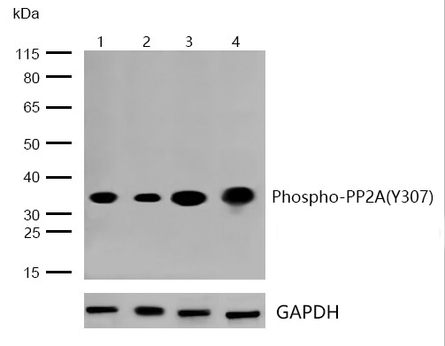
All lanes : PP2A(Phospho-Y307) Rabbit mAb at 1/1k dilution
Lane 1 : A431 whole cell lysates
Lane 2 : F9 whole cell lysates
Lane 3 : PC12 whole cell lysates
Lane 4 : Mouse spleen lysates
Lysates/proteins at 20 µg per lane.
Secondary
All lanes : Goat Anti-Rabbit IgG H&L (HRP) at 1/20000 dilution
Predicted band size: 35 kDa
Observed band size: 35 kDa
Exposure time: 12 seconds
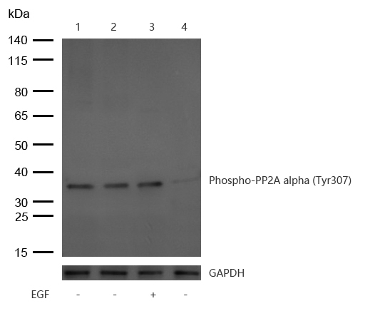
All lanes : PP2A alpha (Phospho-Tyr307) Rabbit mAb at 1/1k dilution
Lane 1 : Rat Kidney lysates
Lane 2 : Mouse Liver lysates
Lane 3 : A431 treated with 100ng/ml EGF for 20min whole cell lysates
Lane 4 : A431 whole cell lysates
Lysates/proteins at 20 µg per lane.
Secondary
All lanes : Goat Anti-Rabbit IgG H&L (HRP) at 1/20000 dilution
Predicted band size: 35 kDa
Observed band size: 35 kDa
Exposure time: 10 seconds
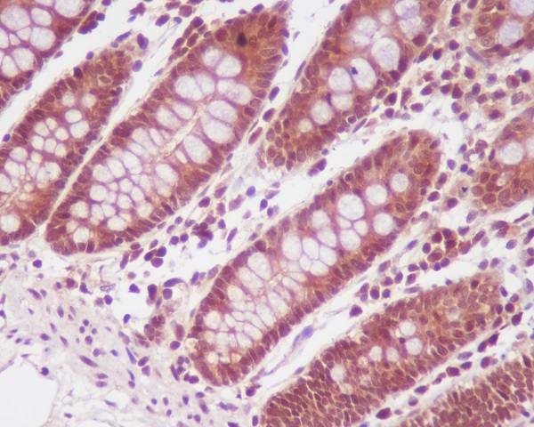
Formalin-fixed;paraffin-embedded human colon tissue stained for PP2A(Phospho-Y307) using 13369 at 1/100 dilution in immunohistochemical analysis.
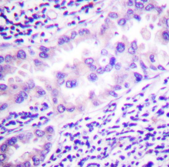
Formalin-fixed;paraffin-embedded human lung carcinoma tissue stained for PP2A(Phospho-Y307) using 13369 at 1/100 dilution in immunohistochemical analysis.
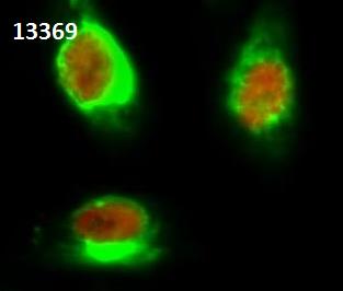
Immunocytochemistry/Immunofluorescence PP2A(Phospho-Y307) antibody (13369)
ICC/IF staining of PP2A(Phospho-Y307) in Hela cells. Cells were fixed with 4% Paraformaldehyde permeabilized with 0.1% Triton X-100.
Samples were incubated with 13369(red) at a working dilution of 1/100.HSP70 Monoclonal antibody (green) was diluted at 1:200.
Goat Anti Rabbit Alexa Fluor 647 was diluted at 1:1000. Goat Anti Mouse Alexa Fluor 488 was diluted at 1:1000.
Protein phosphatase type 2A (PP2A) is an essential protein serine/threonine phosphatase that is conserved in all eukaryotes. PP2A is a key enzyme within various signal transduction pathways as it regulates fundamental cellular activities such as DNA replication, transcription, translation, metabolism, cell cycle progression, cell division, apoptosis and development. The core enzyme consists of catalytic C and regulatory A (or PR65) subunits, with each subunit represented by α and β isoforms. Additional regulatory subunits belong to four different families of unrelated proteins. Both the B (or PR55) and B' regulatory protein families contain α, β, γ and δ isoforms, with the B' family also including an ε protein. B'' family proteins include PR72, PR130, PR59 and PR48 isoforms, while striatin (PR110) and SG2NA (PR93) are both members of the B''' regulatory protein family. These B subunits competitively bind to a shared binding site on the core A subunit. This variable array of holoenzyme components, particularly regulatory B subunits, allows PP2A to act in a diverse set of functions. PP2A function is regulated by expression, localization, holoenzyme composition and post-translational modification. Phosphorylation of PP2A at Tyr307 by Src occurs in response to EGF or insulin and results in a substantial reduction of PP2A activity. Reversible methylation on the carboxyl group of Leu309 of PP2A has been observed. Methylation alters the conformation of PP2A, as well as its localization and association with B regulatory subunits.


 Yes
Yes