产品详情
产品名称Bcl-2 Rabbit mAb
克隆号SZ10-03
来源种属Recombinant Rabbit
克隆性Monoclonal antibody
纯化ProA affinity purified
应用WB, ICC/IF, IHC, IP, FC
种属反应性Human;Mouse
免疫原描述recombinant protein
标记Unconjugated
别名Apoptosis regulator Bcl 2 antibody Apoptosis regulator Bcl-2 antibody Apoptosis regulator Bcl2 antibody AW986256 antibody B cell CLL/lymphoma 2 antibody B cell leukemia/lymphoma 2 antibody Bcl-2 antibody Bcl2 antibody BCL2_HUMAN antibody C430015F12Rik antibody D630044D05Rik antibody D830018M01Rik antibody Leukemia/lymphoma, B-cell, 2 antibody Oncogene B-cell leukemia 2 antibody PPP1R50 antibody Protein phosphatase 1, regulatory subunit 50 antibody
数据库入口号Swiss-Prot#:P10415
Uniprot
P10415
计算分子量26 kDa
实际分子量26 kDa
配方1*TBS (pH7.4), 1%BSA, 40%Glycerol. Preservative: 0.05% Sodium Azide.
保存Store at -20˚C
应用详情
WB: 1:1,000-1:2,000
IHC: 1:50-1:500
ICC: 1:50-1:200
FC: 1:50-1:100
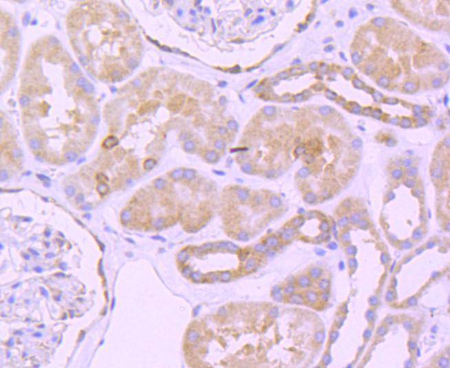
Immunohistochemical analysis of paraffin-embedded human kidney tissue using anti-Bcl-2 antibody. Counter stained with hematoxylin.
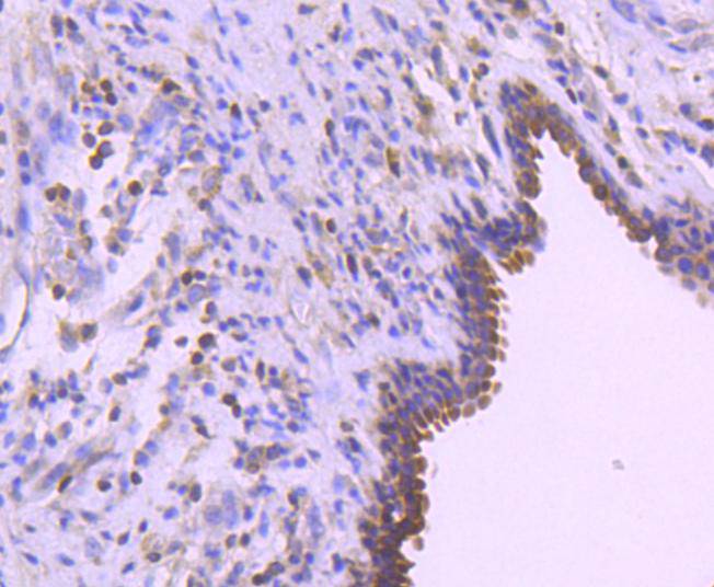
Immunohistochemical analysis of paraffin-embedded human breast carcinoma tissue using anti-Bcl-2 antibody. Counter stained with hematoxylin.
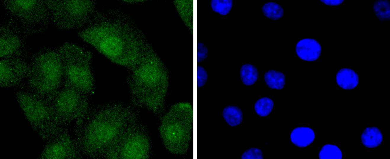
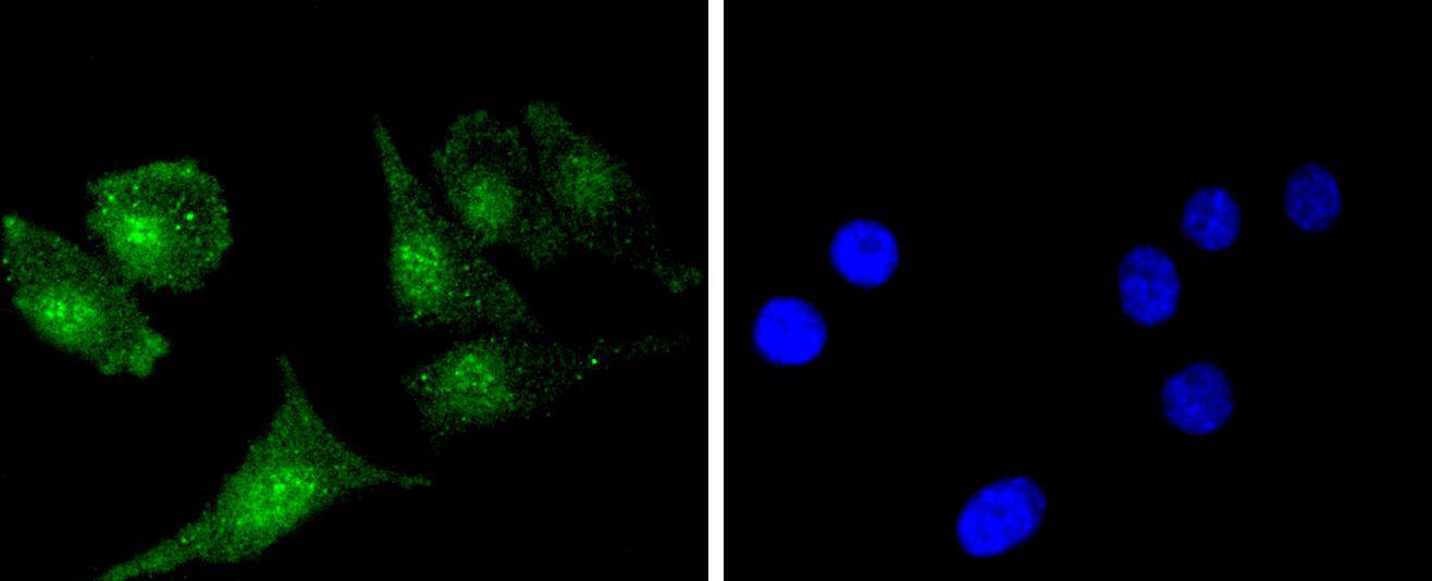
ICC staining Bcl-2 in A549 cells (green). The nuclear counter stain is DAPI (blue). Cells were fixed in paraformaldehyde, permeabilised with 0.25% Triton X100/PBS.
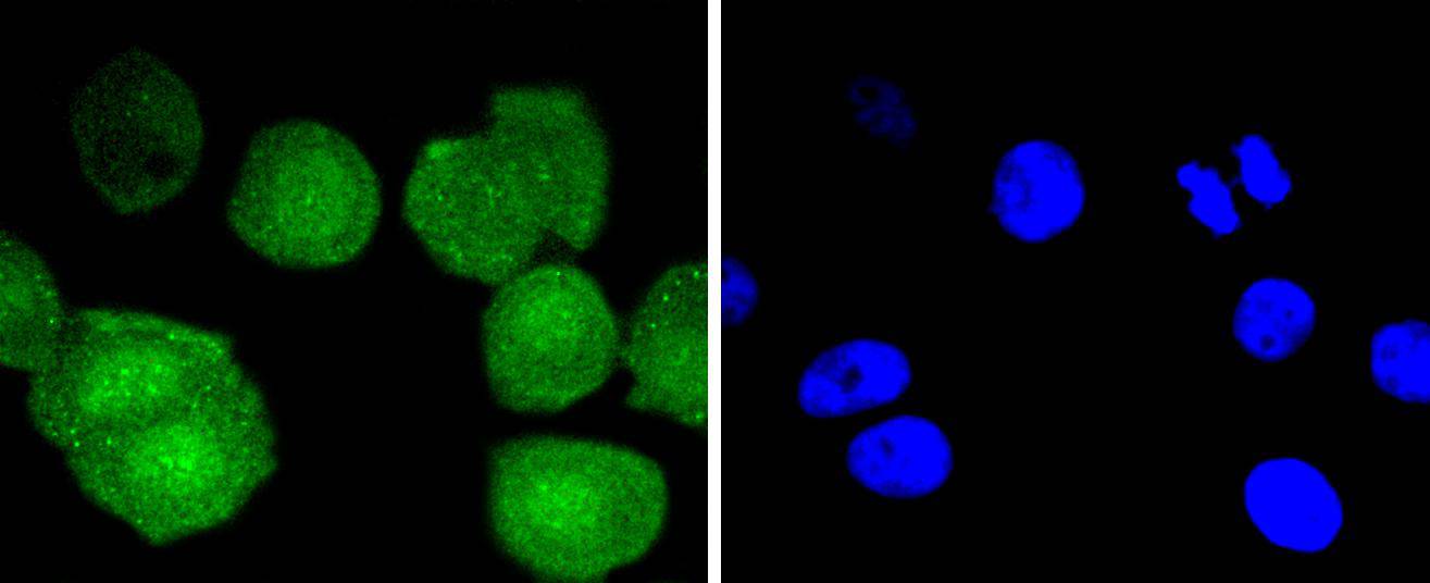
ICC staining Bcl-2 in MCF-7 cells (green). The nuclear counter stain is DAPI (blue). Cells were fixed in paraformaldehyde, permeabilised with 0.25% Triton X100/PBS.
ICC staining Bcl-2 in SH-SY-5Y cells (green). The nuclear counter stain is DAPI (blue). Cells were fixed in paraformaldehyde, permeabilised with 0.25% Triton X100/PBS.
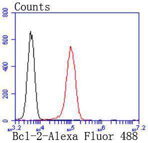
Flow cytometric analysis of Jurkat cells with Bcl-2 antibody at 1/50 dilution (red) compared with an unlabelled control (cells without incubation with primary antibody; black). Alexa Fluor 488-conjugated goat anti rabbit IgG was used as the secondary antibody.
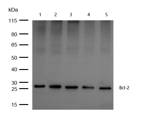
All lanes : Bcl-2 Rabbit mAb at 1/1k dilution
Lane 1 : JK whole cell lysates
Lane 2 : Hela whole cell lysates
Lane 3 : 3T3 whole cell lysates
Lane 4 : Mouse brain lysates whole cell lysatesLane 5 : Mouse lung lysates whole cell lysates
Lysates/proteins at 20 µg per lane.
Secondary
All lanes : Goat Anti-Rabbit IgG H&L (HRP) at 1/20000 dilution
Predicted band size: 26 kDa
Observed band size: 26 kDa
Exposure time: 4 seconds
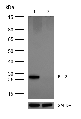
All lanes : Bcl-2 Rabbit mAb at 1/1k dilution
Lane 1 : Wild-type HAP1 cell lysate
Lane 2 : Bcl-2 knockdown HAP1 cell lysate
Lysates/proteins at 20 µg per lane.
Apoptosis is defined as a set of cascades which, when initiated, programs the cell to undergo lethal changes such as membrane blebbing, mitochondrial break down and DNA fragmentation. Bcl-2 is one among many key regulators of apoptosis, which are essential for proper development, tissue homeostasis, and protection against foreign pathogens. Human Bcl-2 is an anti-apoptotic, membrane-associated oncoprotein that can promote cell survival through protein-protein interactions with other Bcl-2 related family members, such as the death suppressors Bcl-xl, Mcl-1, Bcl-w, and A1 or the death agonists Bax, Bak, Bik, Bad, and Bid. The anti-apoptotic function of Bcl-2 can also be regulated through proteolytic processing and phospho-rylation. Bcl-2 may promote cell survival by interfering with the activation of the cytochrome c/Apaf-1 pathway through stabilization of the mitochondrial membrane. Mutations in the Bcl-2 gene can contribute to cancers where normal physiological cell death mechanisms are compromised by deregulation of the anti-apoptotic influence of Bcl-2.
如果您使用该产品48675发表了文章,请通知我们,让我们可以引用您的文献。
et al,LncRNA SERPINB9P1 Mitigates Cerebral Injury Induced by Oxygen‒Glucose Deprivation/Reoxygenation by Interacting with HSPA2.
, (2025),
PMID:
39798045
et al,Transcriptomic Investigation of FoxM1-Mediated Neuroprotection by hAEC-Derived Exosomes in an In Vitro Ischemic Stroke Model
, (2025),
PMID:
41154771


 Yes
Yes