产品详情
产品名称MEK1 Rabbit mAb
克隆号SZ22-01
来源种属Recombinant Rabbit
克隆性 Monoclonal antibody
纯化ProA affinity purified
应用WB, ICC/IF, IHC, IP, FC
种属反应性Hu, Ms, Rt, Cow, Dog
免疫原描述recombinant protein
标记Unconjugated
别名Dual specificity mitogen activated protein kinase kinase 1 antibody Dual specificity mitogen-activated protein kinase kinase 1 antibody ERK activator kinase 1 antibody MAP kinase kinase 1 antibody MAP kinase/Erk kinase 1 antibody MAP2K1 antibody MAPK/ERK kinase 1 antibody MAPKK 1 antibody MAPKK1 antibody MEK 1 antibody Mek1 antibody MEKK1 antibody Mitogen activated protein kinase kinase 1 antibody MKK 1 antibody MKK1 antibody MP2K1_HUMAN antibody PRKMK1 antibody Protein kinase mitogen activated kinase 1 (MAP kinase kinase 1) antibody Protein kinase mitogen activated, kinase 1 antibody
数据库入口号Swiss-Prot#:Q02750
Uniprot
Q02750
计算分子量43 kDa
配方1*TBS (pH7.4), 1%BSA, 40%Glycerol. Preservative: 0.05% Sodium Azide.
保存Store at -20˚C
应用详情
WB: 1:1,000-1:2,000
IHC: 1:50-1:200
ICC: 1:50-1:200
FC: 1:50-1:100
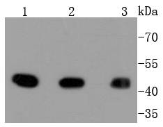
Western blot analysis of MEK1 on different lysates using anti-MEK1 antibody at 1/1,000 dilution. Positive control: Lane 1: A431 Lane 2: HepG2 Lane 3: Hela
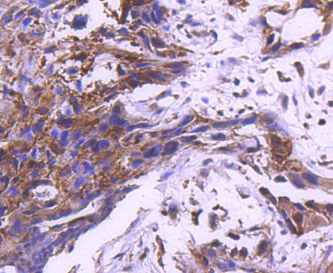
Immunohistochemical analysis of paraffin-embedded human breast carcinoma tissue using anti-MEK1 antibody. Counter stained with hematoxylin.
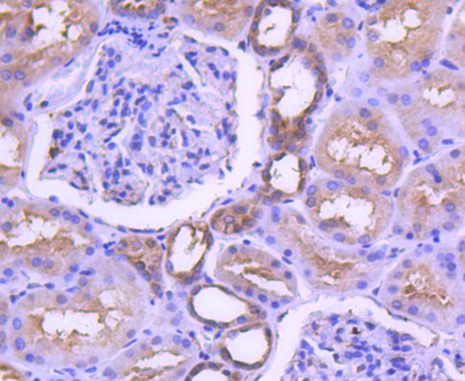
Immunohistochemical analysis of paraffin-embedded human kidney tissue using anti-MEK1 antibody. Counter stained with hematoxylin.
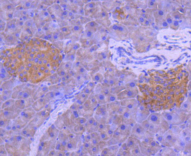
Immunohistochemical analysis of paraffin-embedded mouse pancreas tissue using anti-MEK1 antibody. Counter stained with hematoxylin.
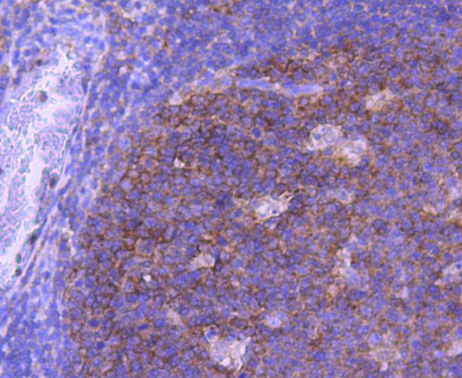
Immunohistochemical analysis of paraffin-embedded human tonsil tissue using anti-MEK1 antibody. Counter stained with hematoxylin.
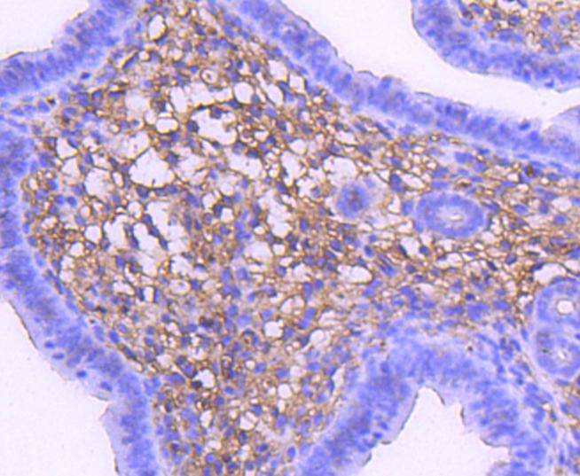
Immunohistochemical analysis of paraffin-embedded mouse uterus tissue using anti-MEK1 antibody. Counter stained with hematoxylin.
ICC staining MEK1 in Hela cells (green). The nuclear counter stain is DAPI (blue). Cells were fixed in paraformaldehyde, permeabilised with 0.25% Triton X100/PBS.
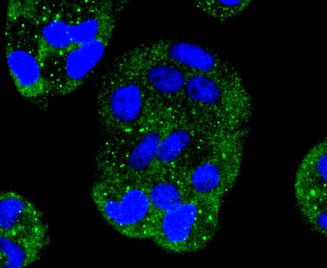
ICC staining MEK1 in MCF-7 cells (green). The nuclear counter stain is DAPI (blue). Cells were fixed in paraformaldehyde, permeabilised with 0.25% Triton X100/PBS.
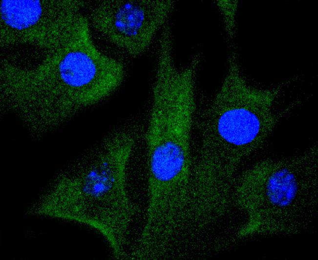
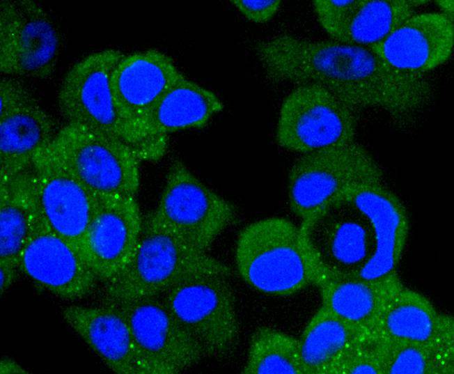
ICC staining MEK1 in NIH/3T3 cells (green). The nuclear counter stain is DAPI (blue). Cells were fixed in paraformaldehyde, permeabilised with 0.25% Triton X100/PBS.
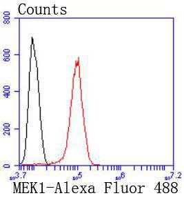
Flow cytometric analysis of Hela cells with MEK1 antibody at 1/50 dilution (red) compared with an unlabelled control (cells without incubation with primary antibody; black). Alexa Fluor 488-conjugated goat anti rabbit IgG was used as the secondary antibody
A family of protein kinases located upstream of the MAP kinases and responsible for their activation has been identified. The prototype member of this family, designated MAP kinase kinase, or MEK-1, specifically phosphorylates the MAP kinase regulatory threonine and tyrosine residues present in the Thr-Glu-Tyr motif of ERK. A second MEK family member, MEK-2, resembles MEK-1 in its substrate specificity. MEK-3 (or MKK-3) functions to activate p38 MAP kinase, and MEK-4 (also called SEK1 or MKK-4) activates both p38 and JNK MAP kinases. MEK-5 appears to specifically phosphorylate ERK5, whereas MEK-6 phosphorylates p38 and p38b. MEK-7 (or MKK-7) phosphorylates and activates the JNK signal transduction pathway.
如果您使用该产品48682发表了文章,请通知我们,让我们可以引用您的文献。