
 Yes
Yes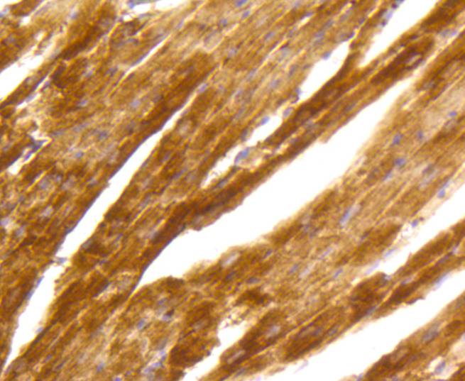
Immunohistochemical analysis of paraffin-embedded mouse heart tissue using anti-PKC alpha antibody. Counter stained with hematoxylin.
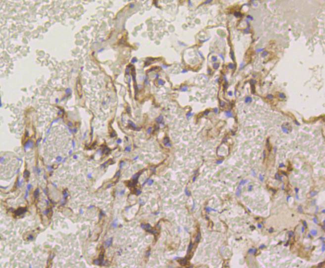
Immunohistochemical analysis of paraffin-embedded human lung tissue using anti-PKC alpha antibody. Counter stained with hematoxylin.
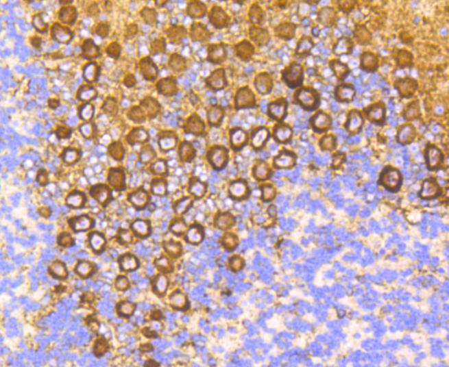
Immunohistochemical analysis of paraffin-embedded mouse brain tissue using anti-PKC alpha antibody. Counter stained with hematoxylin.
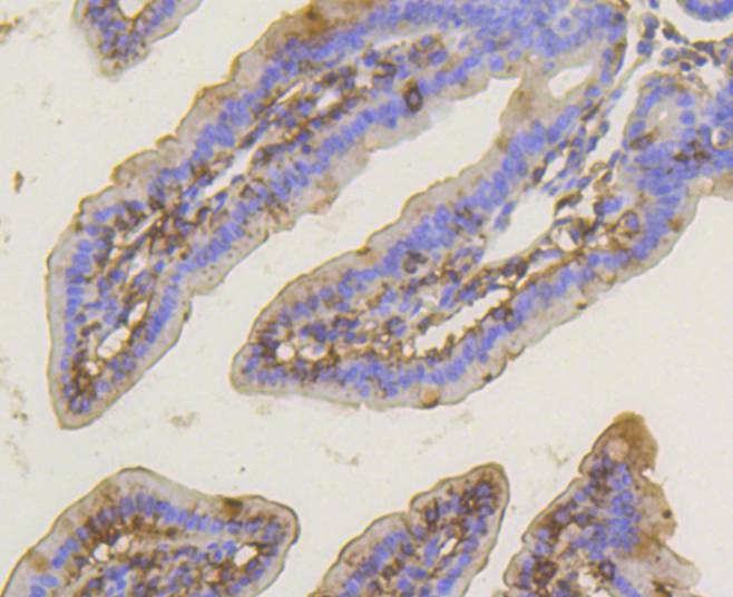
Immunohistochemical analysis of paraffin-embedded mouse small intestine tissue using anti-PKC alpha antibody. Counter stained with hematoxylin.
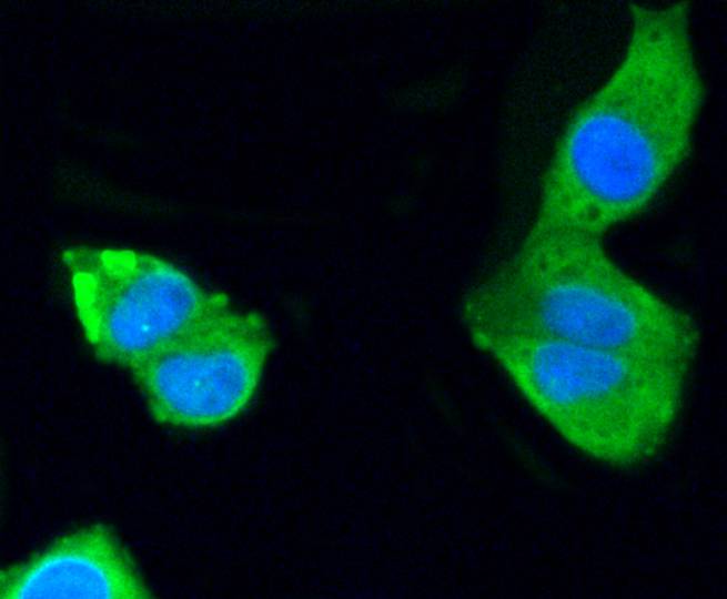
ICC staining PKC alpha in Hela cells (green). The nuclear counter stain is DAPI (blue). Cells were fixed in paraformaldehyde, permeabilised with 0.25% Triton X100/PBS.
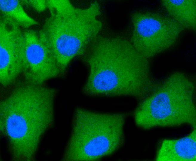
ICC staining PKC alpha in MCF-7 cells (green). The nuclear counter stain is DAPI (blue). Cells were fixed in paraformaldehyde, permeabilised with 0.25% Triton X100/PBS.
ICC staining PKC alpha in CRC cells (green). The nuclear counter stain is DAPI (blue). Cells were fixed in paraformaldehyde, permeabilised with 0.25% Triton X100/PBS.
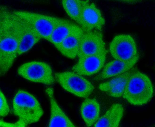
ICC staining PKC alpha in A549 cells (green). The nuclear counter stain is DAPI (blue). Cells were fixed in paraformaldehyde, permeabilised with 0.25% Triton X100/PBS.
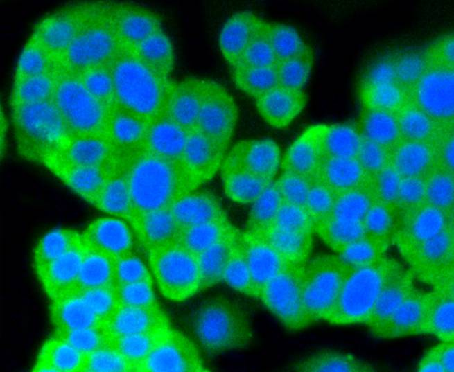
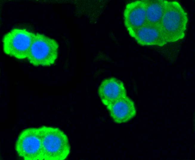
ICC staining PKC alpha in PC12 cells (green). The nuclear counter stain is DAPI (blue). Cells were fixed in paraformaldehyde, permeabilised with 0.25% Triton X100/PBS.
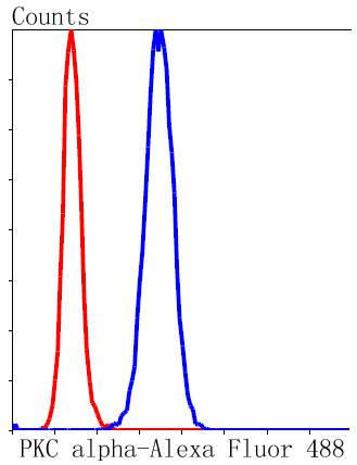
Flow cytometric analysis of Hela cells with PKC alpha antibody at 1/50 dilution (blue) compared with an unlabelled control (cells without incubation with primary antibody; red). Alexa Fluor 488-conjugated goat anti rabbit IgG was used as the secondary antibody.
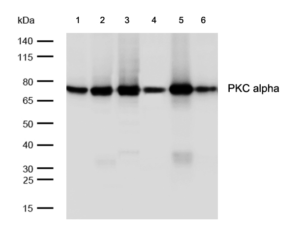
All lanes: PKC alpha Rabbit mAb at 1/1k dilutionLane 1 : Hela whole cell lysates Lane 2 : 293 whole cell lysates Lane 3 : C6 whole cell lysates Lane 4 : Mouse liver lysates Lane 5 : Mouse spleen lysates Lane 6 : Rat liver lysates Lysates/proteins at 20 µg per lane.SecondaryAll lanes : Goat Anti-Rabbit IgG H&L (HRP) at 1/20000 dilutionPredicted band size: 77 kDa Observed band size: 75 kDaExposure time: 5 seconds
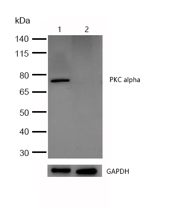
All lanes :PKC alpha Rabbit mAb at 1/1k dilutionLane 1 : Wild-type HAP1 cell lysateLane 2 : PKC alpha knockdown HAP1 cell lysateLysates/proteins at 20 µg per lane.