产品详情
产品名称p63 Rabbit mAb
克隆号SC06-31
来源种属Recombinant Rabbit
克隆性 Monoclonal antibody
纯化ProA affinity purified
应用WB, ICC/IF, IHC, FC
种属反应性Human;Mouse;Rat
免疫原描述recombinant protein
标记Unconjugated
别名AIS antibody Amplified in squamous cell carcinoma antibody B(p51A) antibody B(p51B) antibody Chronic ulcerative stomatitis protein antibody CUSP antibody DN p63 alpha 1 antibody DNp63 antibody EEC3 antibody id:ibd3516 antibody Keratinocyte transcription factor antibody Keratinocyte transcription factor KET antibody KET antibody LMS antibody MGC115972 antibody MGC192897 antibody NBP antibody OFC8 antibody OTTHUMP00000209732 antibody OTTHUMP00000209733 antibody OTTHUMP00000209734 antibody OTTHUMP00000209735 antibody OTTHUMP00000209737 antibody OTTHUMP00000209738 antibody OTTHUMP00000209739 antibody OTTHUMP00000209740 antibody OTTHUMP00000209741 antibody OTTHUMP00000209742 antibody OTTHUMP00000209743 antibody OTTHUMP00000209744 antibody p40 antibody p51 antibody P51/P63 antibody p53-related protein p63 antibody p53CP antibody p63 antibody P63_HUMAN antibody p73H antibody p73L antibody RHS antibody SHFM4 antibody TAp63alpha antibody TP53CP antibody TP53L antibody TP63 antibody TP73L antibody Transformation related protein 63 antibody Transformation-related protein 63 antibody Trp53rp1 antibody Trp63 antibody Tumor protein 63 antibody Tumor protein p53-competing protein antibody Tumor protein p53-like antibody Tumor protein p63 antibody Tumor protein p63 deltaN isoform delta antibody Tumor protein p73 antibody Tumor protein p73-like antibody
数据库入口号Swiss-Prot#:Q9H3D4
Uniprot
Q9H3D4
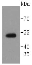
计算分子量55 kDa
配方1*TBS (pH7.4), 1%BSA, 40%Glycerol. Preservative: 0.05% Sodium Azide.
保存Store at -20˚C
应用详情
WB: 1:1,000-1:2,000
IHC: 1:50-1:200
ICC: 1:50-1:200
FC: 1:50-1:100
Western blot analysis of p63 on human kidney lysates using anti-p63 antibody at 1/1,000 dilution.
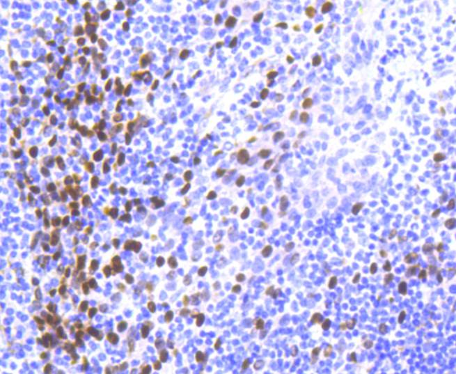
Immunohistochemical analysis of paraffin-embedded human tonsil tissue using anti-p63 antibody. Counter stained with hematoxylin.
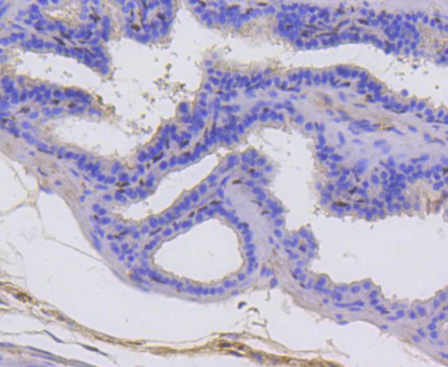
Immunohistochemical analysis of paraffin-embedded mouse prostate tissue using anti-p63 antibody. Counter stained with hematoxylin.
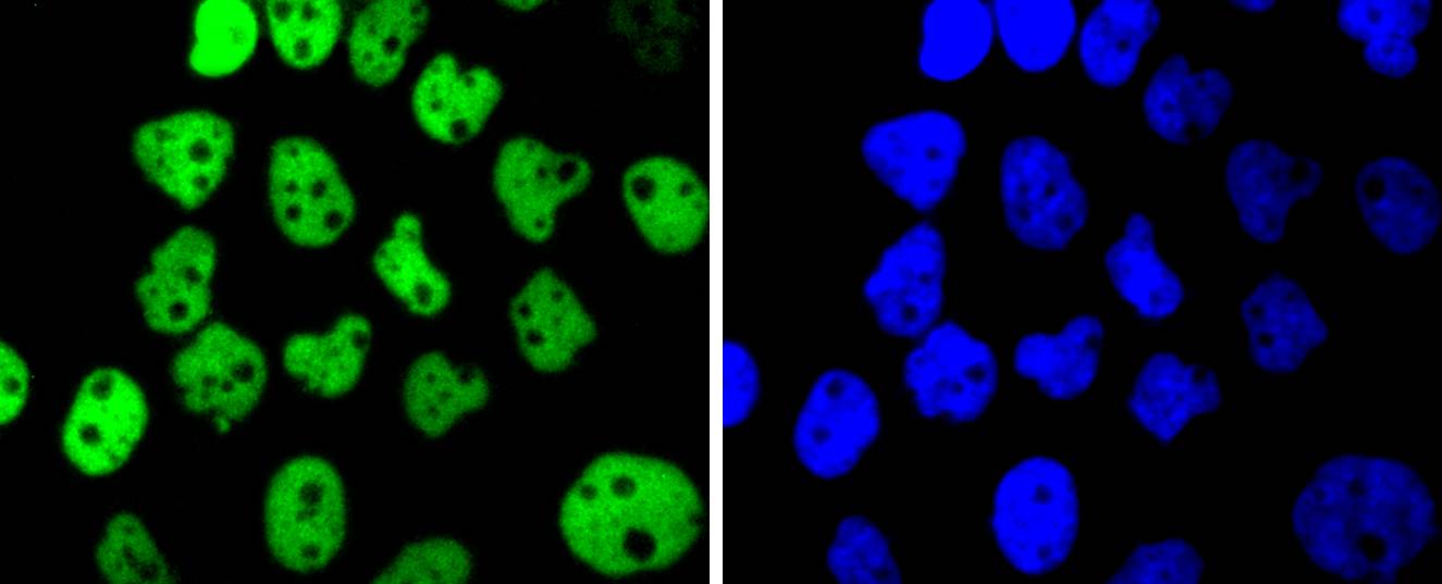
ICC staining p63 in A431 cells (green). The nuclear counter stain is DAPI (blue). Cells were fixed in paraformaldehyde, permeabilised with 0.25% Triton X100/PBS.
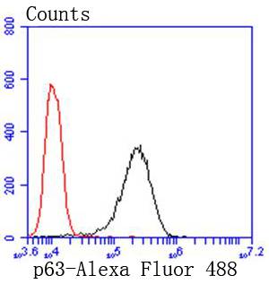
Flow cytometric analysis of A431 cells with p63 antibody at 1/50 dilution (black) compared with an unlabelled control (cells without incubation with primary antibody; red). Alexa Fluor 488-conjugated goat anti rabbit IgG was used as the secondary antibody.
The p53 tumor suppressor protein plays a major role in cellular response to DNA damage and other genomic aberrations. Activation of p53 can lead to either cell cycle arrest and DNA repair or apoptosis. In addition to p53, mammalian cells contain two p53 family members, p63 and p73, which are similar to p53 in both structure and function. While p63 can induce p53-responsive genes and apoptosis, mutation of p63 rarely results in tumors. Research investigators frequently observe amplification of the p63 gene in squamous cell carcinomas of the lung, head and neck. The p63 gene contains an alternative transcription initiation site that yields a 40 kDa δNp63 lacking the transactivation domain, and alternative splicing at the carboxy-terminus yields the α, β, and γ isoforms.
如果您使用该产品48958发表了文章,请通知我们,让我们可以引用您的文献。


 Yes
Yes