产品详情
产品名称Apg7 Rabbit mAb
克隆号SC06-30
来源种属Recombinant Rabbit
克隆性 Monoclonal antibody
纯化ProA affinity purified
应用WB, ICC/IF, IHC, IP, FC
种属反应性Human
免疫原描述recombinant protein
标记Unconjugated
别名1810013K23Rik antibody Apg 7 antibody APG7 autophagy 7 like antibody APG7 autophagy 7-like (S. cerevisiae) antibody APG7 like antibody APG7, S. cerevisiae, homolog of antibody APG7-like antibody APG7L antibody ATG 7 antibody ATG12-activating enzyme E1 ATG7 antibody ATG7 antibody ATG7 autophagy related 7 homolog (S. cerevisiae) antibody ATG7 autophagy related 7 homolog antibody ATG7_HUMAN antibody Atg7l antibody Autophagy 7, S. cerevisiae, homolog of antibody Autophagy related protein 7 antibody Autophagy-related 7 (yeast) antibody Autophagy-related protein 7 antibody DKFZp434N0735 antibody GSA 7 antibody GSA7 antibody hAGP7 antibody Ubiquitin activating enzyme E1 like protein antibody Ubiquitin-activating enzyme E1-like protein antibody Ubiquitin-like modifier-activating enzyme ATG7 antibody
数据库入口号Swiss-Prot#:O95352
Uniprot
O95352
计算分子量78 kDa
配方1*TBS (pH7.4), 1%BSA, 40%Glycerol. Preservative: 0.05% Sodium Azide.
保存Store at -20˚C
应用详情
WB: 1:1,000-1:2,000
IHC: 1:50-1:200
ICC: 1:50-1:200
FC: 1:50-1:100
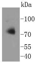
Western blot analysis of Apg7 on Jurkat cells lysates using anti-Apg7 antibody at 1/1,000 dilution.
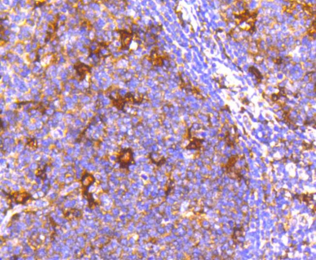
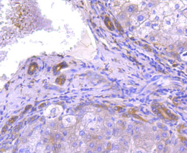
Immunohistochemical analysis of paraffin-embedded human tonsil tissue using anti-Apg7 antibody. Counter stained with hematoxylin.
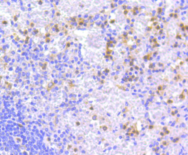
Immunohistochemical analysis of paraffin-embedded human spleen tissue using anti-Apg7 antibody. Counter stained with hematoxylin.
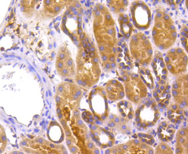
Immunohistochemical analysis of paraffin-embedded human kidney tissue using anti-Apg7 antibody. Counter stained with hematoxylin.
Immunohistochemical analysis of paraffin-embedded mouse brain tissue using anti-Apg7 antibody. Counter stained with hematoxylin.
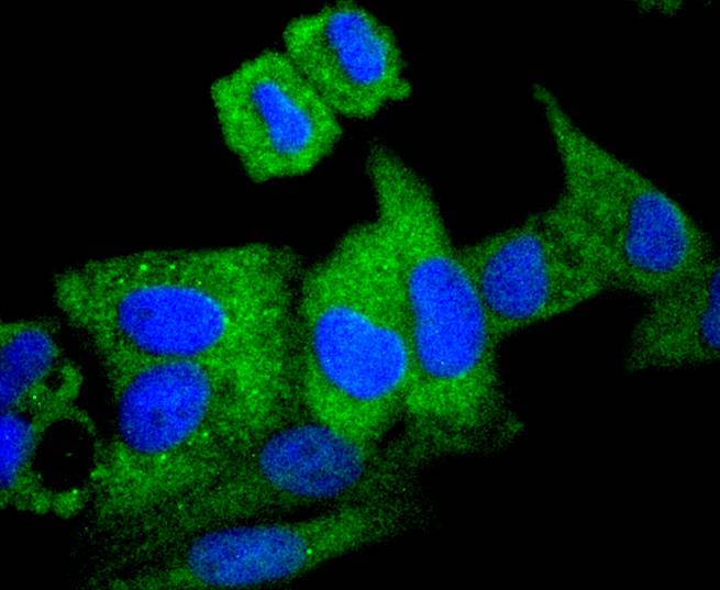
ICC staining Apg7 in Hela cells (green). The nuclear counter stain is DAPI (blue). Cells were fixed in paraformaldehyde, permeabilised with 0.25% Triton X100/PBS.
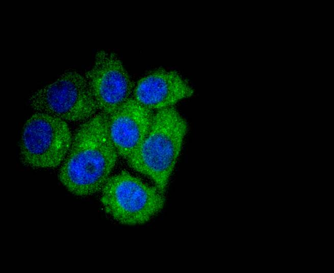
ICC staining Apg7 in HepG2 cells (green). The nuclear counter stain is DAPI (blue). Cells were fixed in paraformaldehyde, permeabilised with 0.25% Triton X100/PBS.
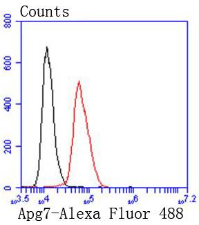
Flow cytometric analysis of Hela cells with Apg7 antibody at 1/50 dilution (red) compared with an unlabelled control (cells without incubation with primary antibody; black). Alexa Fluor 488-conjugated goat anti rabbit IgG was used as the secondary antibody.
In yeast, autophagy is an essential process for survival during nutrient starvation and cell differentiation. The process of autophagy is characterized as a non-selective degradation of cytoplasmic proteins into membrane stuctures called autophagosomes, and it is dependent on several proteins, including the autophagy proteins APG5 and APG7. Yeast Apg7 and the human homolog, APG7, share similarities with the ubiquitin-activating enzyme E1 in Saccharomyces cerevisiae, and are likewise responsible for enzymatically activating the autophagy conjugation system. Apg5 and the human homolog, APG5 (also designated apoptosis specific protein or APS), function as substrates for the autophagy protein APG12. These proteins are covalently bonded together to form APG12/APG5 conjugates, which are required for the progression of autophagy.
如果您使用该产品48968发表了文章,请通知我们,让我们可以引用您的文献。


 Yes
Yes