
 Yes
Yes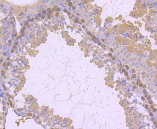
Immunohistochemical analysis of paraffin-embedded mouse testis tissue using anti-PKC delta antibody. Counter stained with hematoxylin.
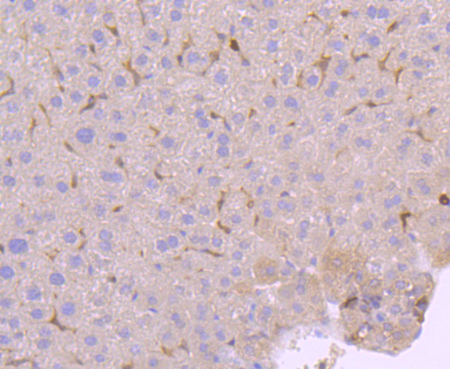
Immunohistochemical analysis of paraffin-embedded mouse liver tissue using anti-PKC delta antibody. Counter stained with hematoxylin.
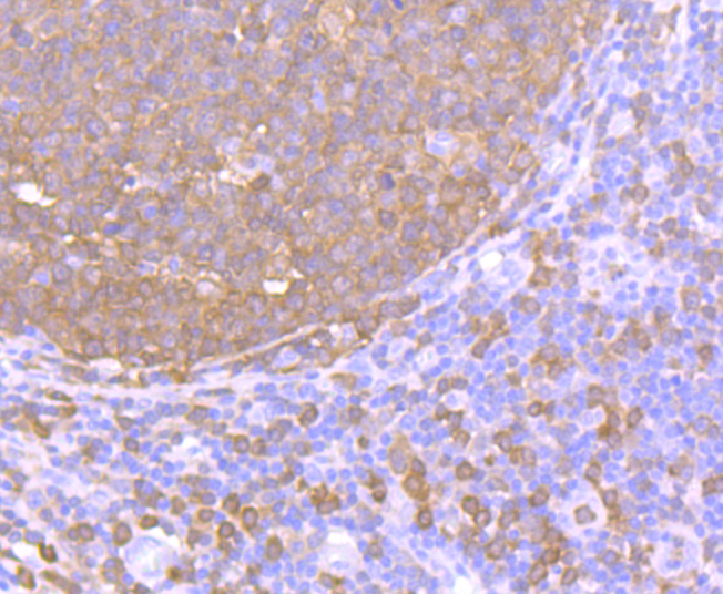
Immunohistochemical analysis of paraffin-embedded human tonsil tissue using anti-PKC delta antibody. Counter stained with hematoxylin.
Immunohistochemical analysis of paraffin-embedded human liver tissue using anti-PKC delta antibody. Counter stained with hematoxylin.
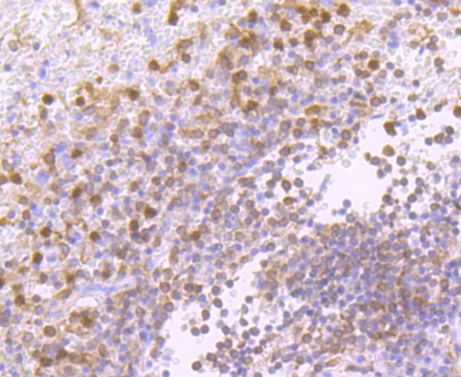
Immunohistochemical analysis of paraffin-embedded human spleen tissue using anti-PKC delta antibody. Counter stained with hematoxylin.
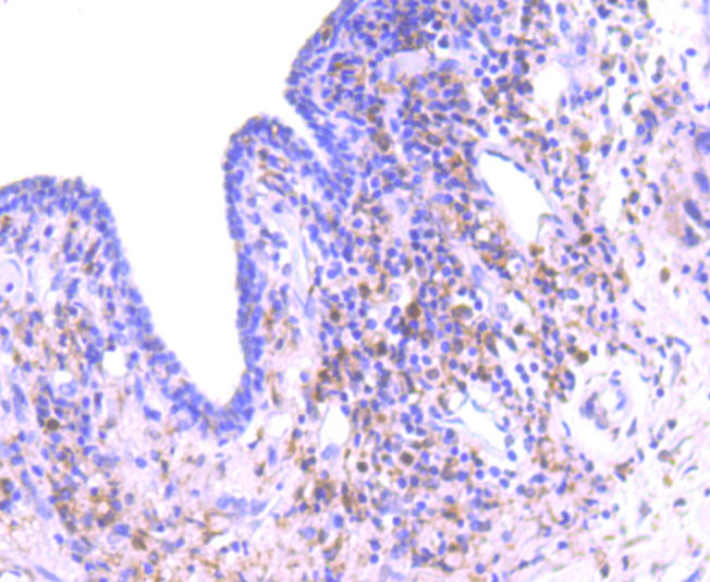
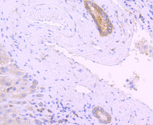
Immunohistochemical analysis of paraffin-embedded human breast carcinoma tissue using anti-PKC delta antibody. Counter stained with hematoxylin.
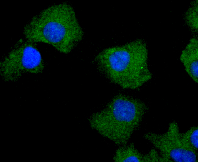
ICC staining PKC delta in A549 cells (green). The nuclear counter stain is DAPI (blue). Cells were fixed in paraformaldehyde, permeabilised with 0.25% Triton X100/PBS.
ICC staining PKC delta in MCF-7 cells (green). The nuclear counter stain is DAPI (blue). Cells were fixed in paraformaldehyde, permeabilised with 0.25% Triton X100/PBS.
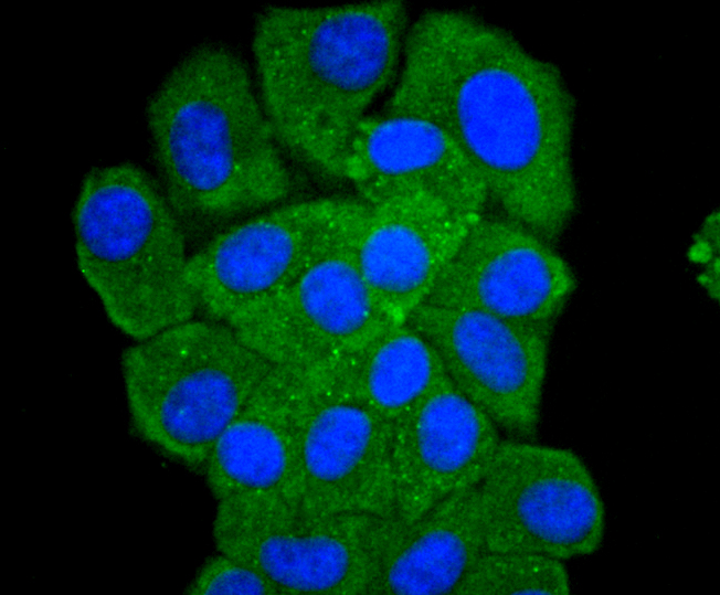
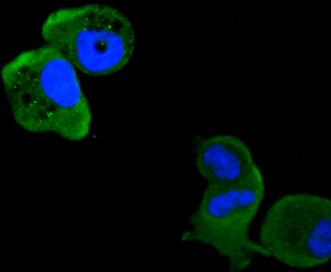
ICC staining PKC delta in HepG2 cells (green). The nuclear counter stain is DAPI (blue). Cells were fixed in paraformaldehyde, permeabilised with 0.25% Triton X100/PBS.
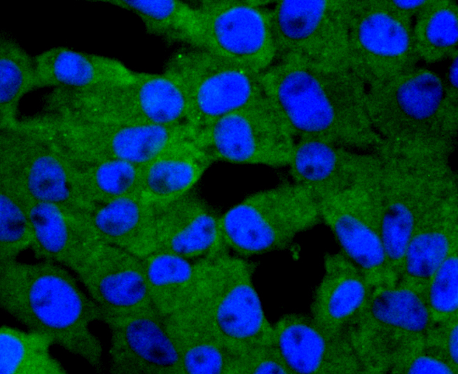
ICC staining PKC delta in RH-35 cells (green). The nuclear counter stain is DAPI (blue). Cells were fixed in paraformaldehyde, permeabilised with 0.25% Triton X100/PBS.
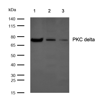
All lanes: PKC delta Rabbit mAb at 1/1k dilutionLane 1 : HeLa whole cell lysates Lane 2 : Mouse brain lysates Lane 3 : Rat brain lysates Lysates/proteins at 20 µg per lane.SecondaryAll lanes : Goat Anti-Rabbit IgG H&L (HRP) at 1/20000 dilutionPredicted band size: 78 kDa Observed band size: 78 kDaExposure time: 6 seconds
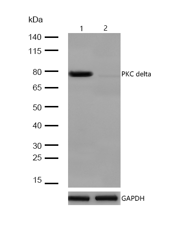
All lanes:PKC delta Rabbit mAb at 1/1k dilutionLane 1 : Wild-type Hela cell lysateLane 2 :PKC delta Rabbit mAb knockdown Hela cell lysateLysates/proteins at 20 µg per lane.