
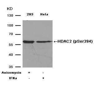
-
JAK-STAT Pathway
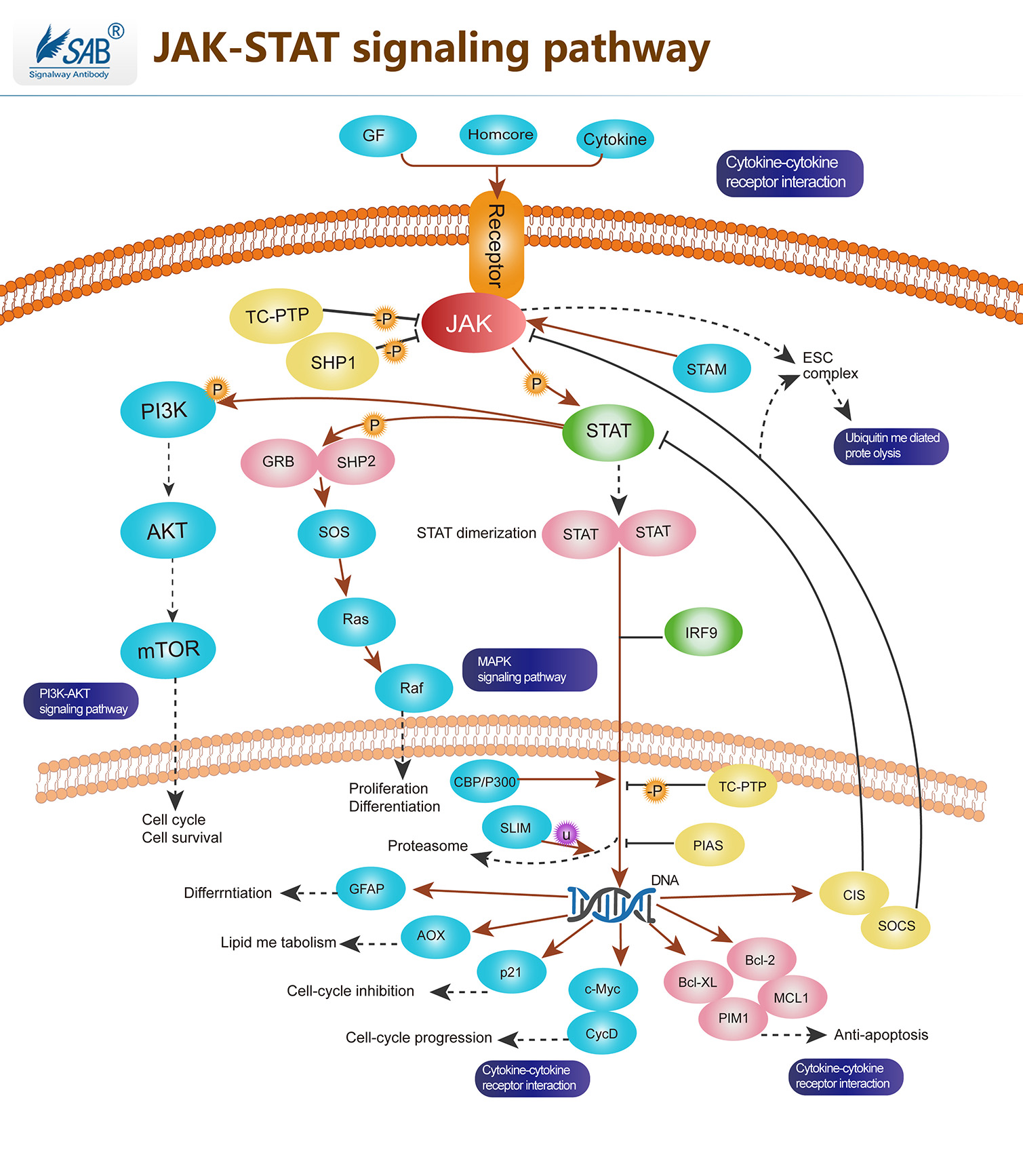
JAK -STAT途径是多种细胞因子和生长因子的主要信号传导机制。JAK激活刺激细胞增殖,分化,细胞迁移和凋亡。这些细胞事件对于造血,免疫发育,乳腺发育和泌乳,脂肪形成,两性性生长和其他过程至关重要。JAK(janus kinase)是一类非受体酪氨酸激酶家族,已发现四个成员,即Jak1 、Jak2 、Jak3 和Tyk2。JAK的N端结构域与受体相结合,C端为激酶结构域。每种激酶成员与特异的细胞因子受体结合。JAK的底物为STAT,即信号转导子和转录激活子(signal transducer and activator of transcription,STAT),N端具有SH2结构域和核定位信号(NLS),中间为DNA结合域,C端有保守的,对其活化至关重要的酪氨酸残基。共发现7个STAT家族成员,分别命名为STAT1至STAT7。STAT被JAK磷酸化后发生二聚化,然后穿过核膜进入核内调节相关基因的表达,这条信号通路称为JAK-STAT途径。
-
Autophagy Pathway
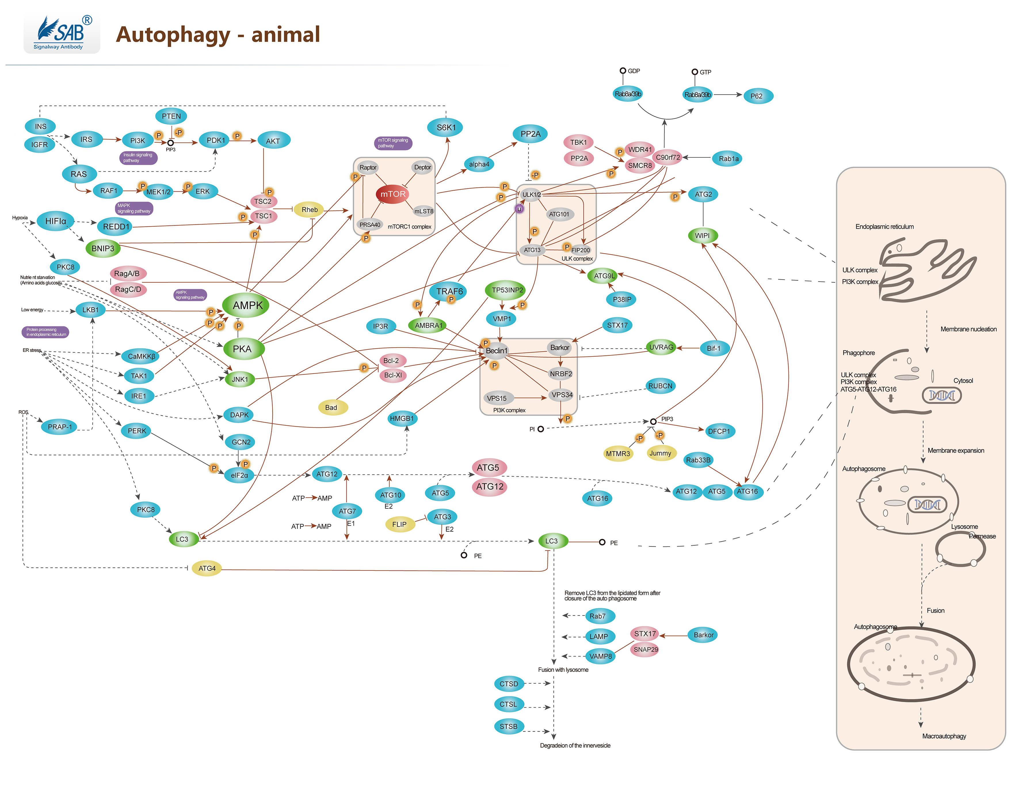
自噬 Autophagy,或称自体吞噬是一个涉及到细胞自身结构通过溶酶体机制,负责将受损的细胞器、错误折叠的蛋白及其他大分子物质等运送至溶酶体降解并再利用的进化保守过程。自噬是广泛存在于真核细胞的现象,并且可分为巨自噬、微自噬和分子伴侣介导的自噬三大类。这是一个受到紧密调控的步骤,此步骤是细胞生长、发育与稳态中的常规步骤,帮助细胞产物在合成、降解以及接下来的循环中保持一个平衡状态。目前已有多份研究表明自噬在许多细胞的分化进程中被不同程度地激活,例如参与血管生成、成骨分化、脂肪生成、神经发生等过程。自噬效应的发生取决于自噬流过程是否完成,而自噬流的意思是自噬的完整动态过程,包括自噬体形成、自噬体与溶酶体融合及后续内含物的降解和回收。
-
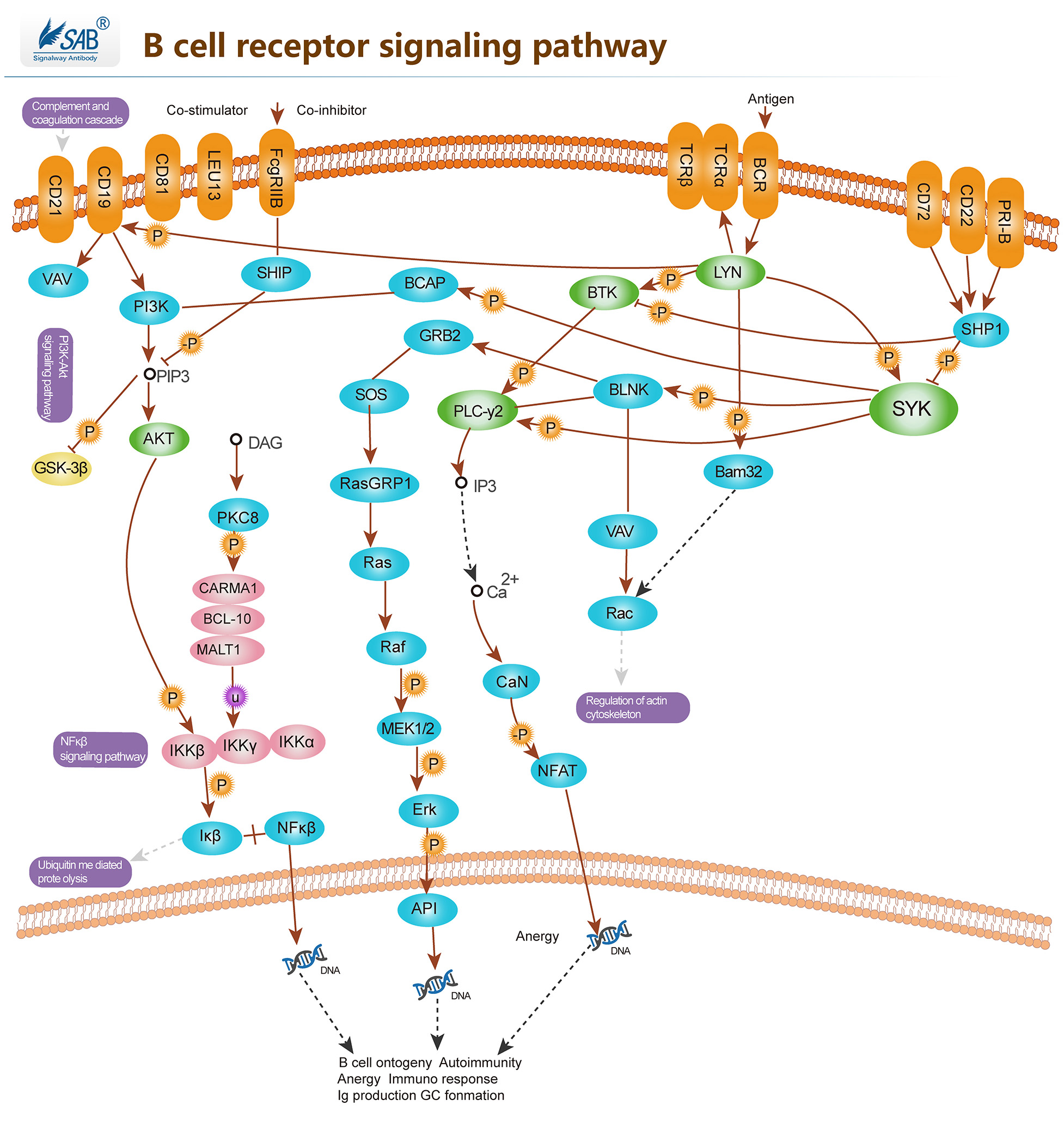
B CELL Pathway
-
Notch signaling pathway
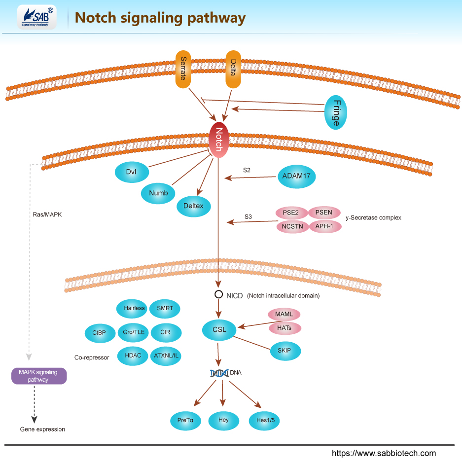
Notch信号通路由Notch受体、Notch配体(DSL蛋白)、CSL (CBF-1,Suppressor of hairless,Lag的合称)DNA结合蛋白、其他的效应物和Notch的调节分子等组成。1917年,Morgan及其同事在突变的果蝇中发现Notch基因,因该基因的部分功能缺失会在果蝇翅膀的边缘造成缺刻(Notch)而得名。哺乳动物有4种Notch受体(Notch1- 4)和5种Notch配体(Delta-like 1, 3, 4,Jagged1和Jagged2)。Notch信号的产生是通过相邻细胞的Notch配体与受体相互作用,Notch蛋白经过三次剪切,由胞内段(NICD)释放入胞质,并进入细胞核与转录因子CSL结合, 形成NICD/CSL转录激活复合体,从而激活HES、HEY、HERP等碱性-螺旋-环-螺旋(basic-helix-loop- helix,bHLH)转录抑制因子家族的靶基因,发挥生物学作用。
-
NF-kappa B signaling pathway
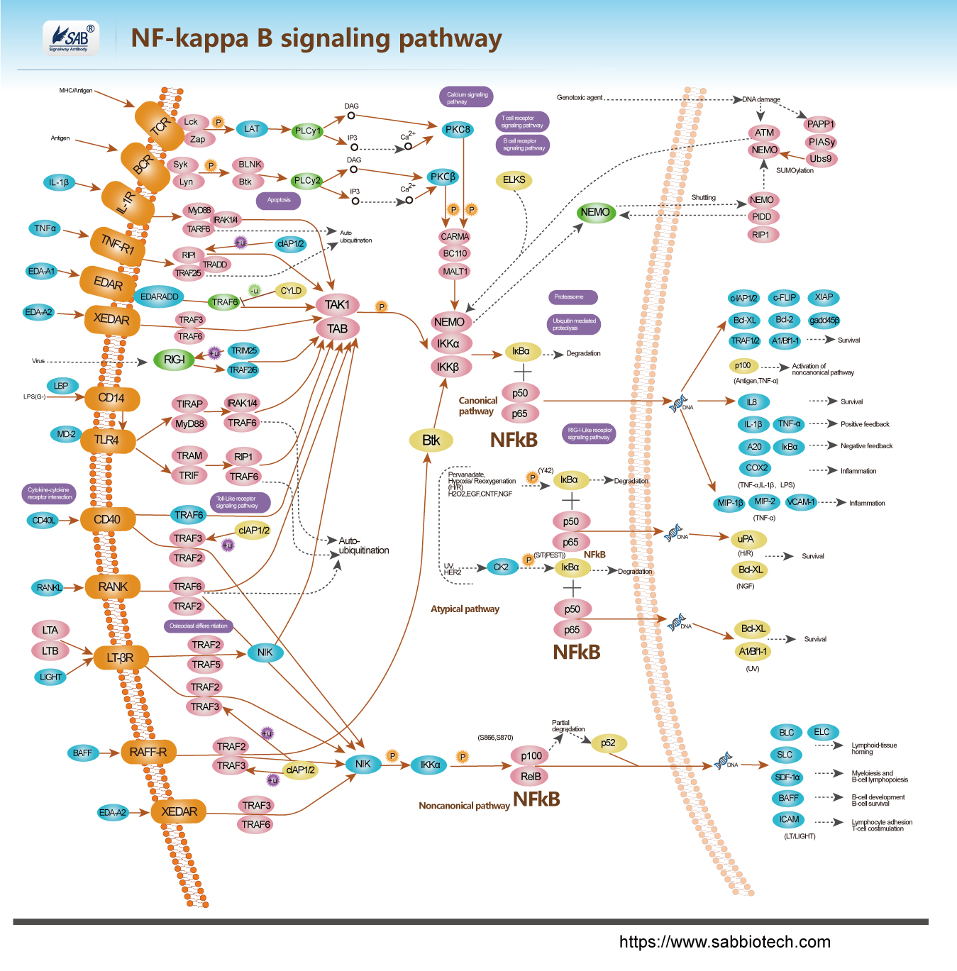
Pathway description:
NF-Kappa B (Nuclear Factor-Kappa B) is a heterodimeric protein composed of different combinations of members of the Rel family of transcription factors. The Rel/ NF-Kappa B family of transcription factors are involved mainly in stress-induced, immune, and inflammatory responses. In addition, these molecules play important roles during the development of certain hemopoietic cells, keratinocytes, and lymphoid organ structures. NF-Kappa B is also an important regulator in cell fate decision, such as programmed cell death and proliferation controlled, and is critical in tumorigenesis. NF-Kappa B is composed of homo- and heterodimers of five members of the Rel family including NF-Kappa B1, NF-Kappa B2,RelA,RelB,and c-Rel. NF-Kappa B can be activated by exposure of cells to LPS or inflammatory cytokines such as TNF or IL-1,growth factors, lymphokines, oxidant-free radical, inhaled particles, viral infection or expression of certain viral or bacterial gene products, UV irradiation, B or T-cell activation, and by other physiological and non physiological stimulin.
Selected Reviews:
Baldwin AS Jr. (1996) The NF-Kappa B and I kappa B proteins:new discoveries and insights.Annu Rev Immunol.14,649.
Vigo Heissmeyer and Daniel Krappmann,et al. (2001)Shared Pathways of I B Kinase-Induced SCF TrCP-Mediated Ubiquitination and Degradation for the NF-B Precursor precursor precursor p105 and I Bacterial. Molecular and Cellular Biology.21,1024.
Joel L,Pomerantz and David Baltimore.(2002)Two pathway to NF-Kappa B.Molecular Cell.10,693.
-
AMPK signaling pathway
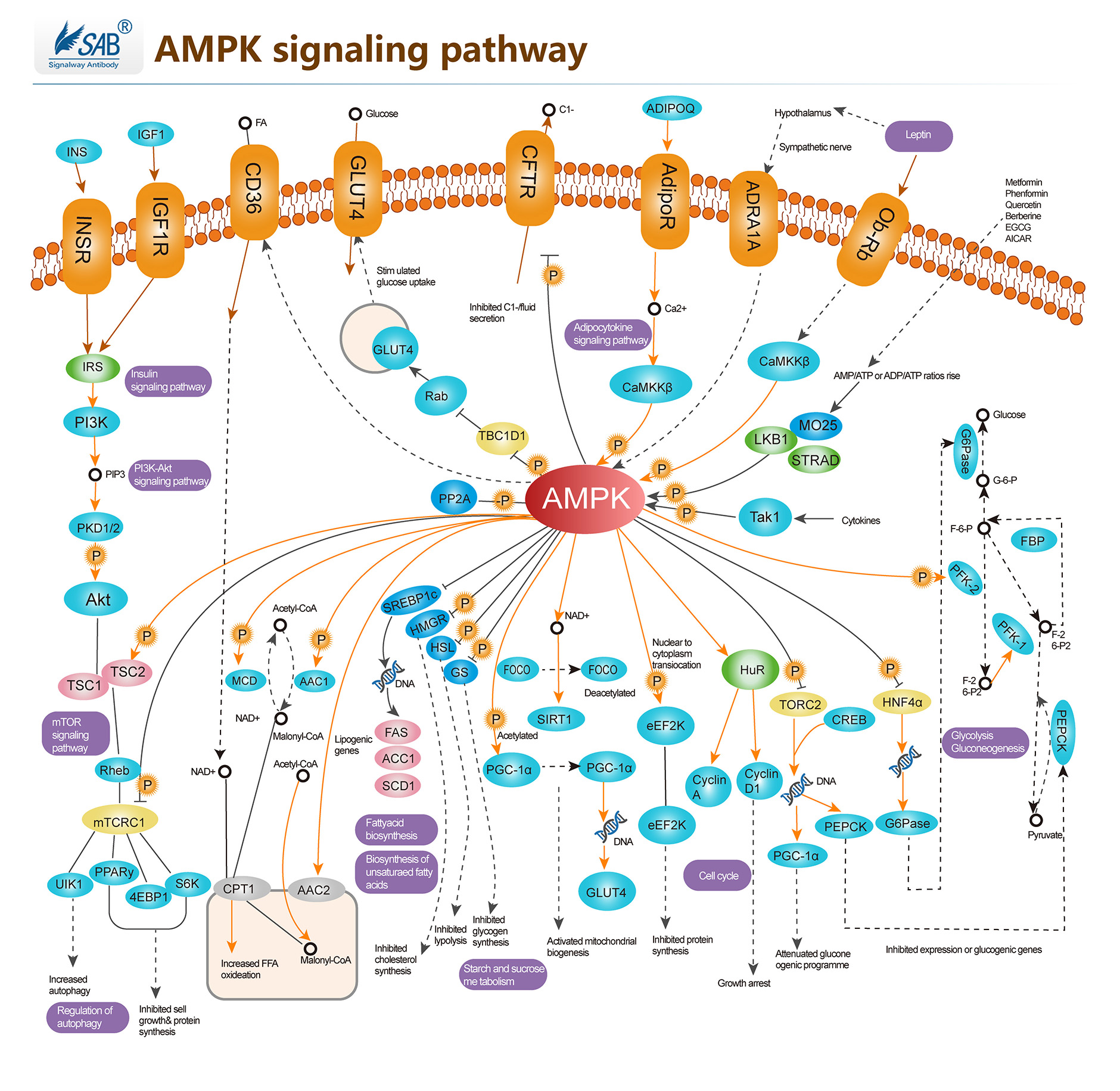
AMPK(AMP-activated protein kinase)信号通路是一种细胞内的信号通路,参与细胞代谢的调节,与细胞的能量代谢密切相关。AMPK是一种重要的蛋白激酶,由蛋白激酶激活蛋白激酶(LKB1)激活,能够响应细胞的能量状态和代谢状态,调节细胞的代谢和生长。AMPK信号通路的主要功能是维持细胞能量平衡,调节葡萄糖和脂肪酸代谢。AMPK活化能够刺激葡萄糖的摄入和运输,促进葡萄糖的利用,同时缺乏能量供应时,AMPK能够促进脂肪酸的氧化,以维持细胞能量平衡。AMPK的活化还能够促进蛋白质的合成,细胞周期的调节,促进细胞的生长和增殖,以维持细胞的稳态。此外,AMPK的活化还与其他相关的信号通路进行调节,如PI3K信号通路、mTOR信号通路、Wnt信号通路等。AMPK的活化可能对治疗糖尿病、肥胖等疾病具有重要的潜在治疗作用。
-
Hedgehog signaling pathway
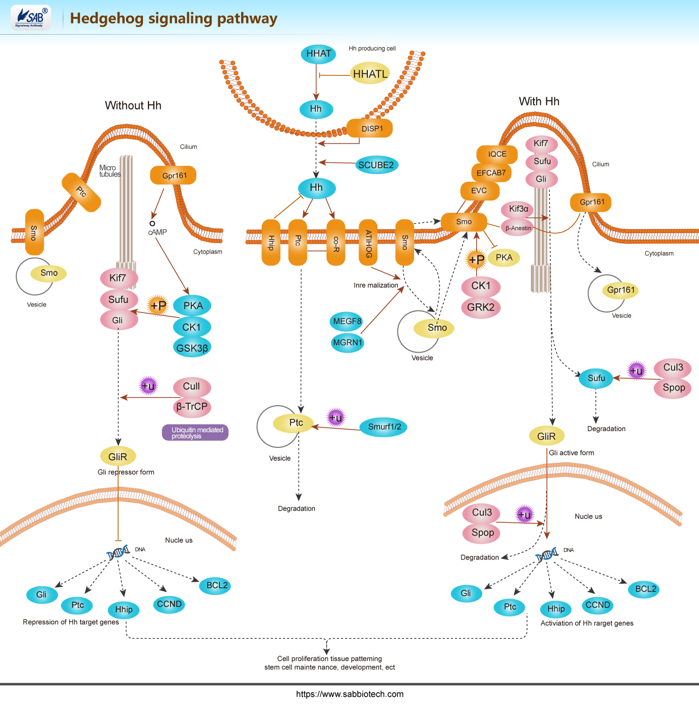
Hh信号传递受靶细胞膜上两种受体Patched(Ptc)和Smoothened(Smo)的控制。受体Ptc由肿瘤抑制基因Patched编码,是由12个跨膜区的单一肽链构成,能与配体直接结合,对Hh信号起负调控作用。受体Smo由原癌基因Smothened 编码,与G蛋白偶联受体同源,由7个跨膜区的单一肽链构成,N端位于细胞外,C端位于细胞内,跨膜区氨基酸序列高度保守,C 末端的丝氨酸与苏氨酸残基为磷酸化部位,蛋白激酶催化时结合磷酸基团。该蛋白家族成员只有当维持全长时才有转录启动子的功能,启动下游靶基因的转录;当羧 基端被蛋白酶体水解后,就形成转录抑制子,抑制下游靶基因的转录。Smo是Hh信号传递所必须的受体。在无Hh、Ptc的情况下,激活Smo可导 致Hh 靶基因的活化;基因Smo突变时,可出现与Hh 基因突变相同的表征。
-
Akt signaling pathway
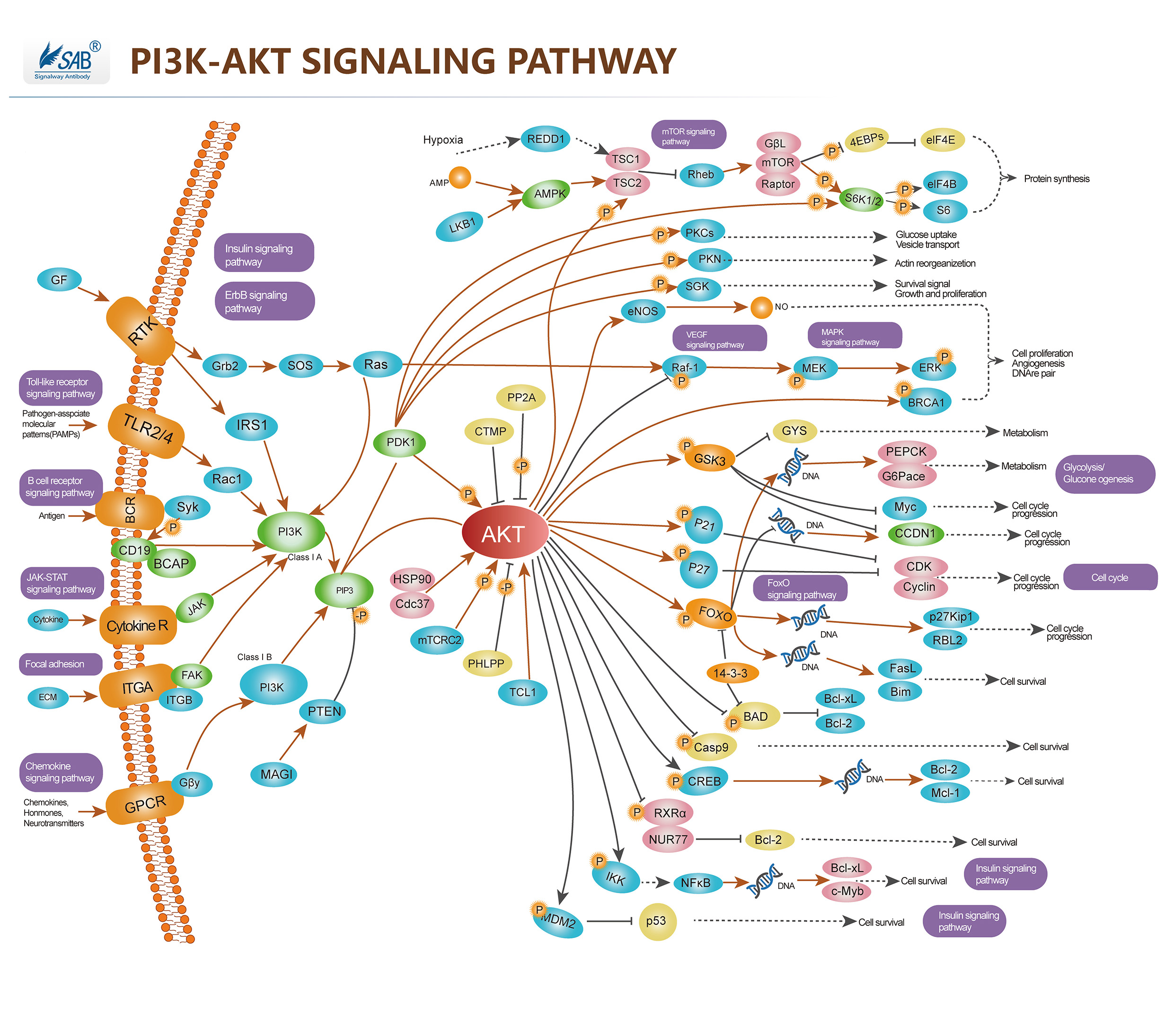
Pathway description:
Akt (v-Akt Murine Thymoma Viral Oncogene)/ PKB (Protein Kinase-B) is a Serine/threonine Kinase that is involved in mediating various biological responses, by phospho-rylation of a number of intracellular proteins, regulates different cellular processes, such as cell growth, cell cycle, apoptosis and glucose metabolism. Akt is activated by several hormones including insulin, growth factors, by signals derived from receptors for extracellular matrix molecules such as integrins, by several forms of cellular stress such as oxidative stress or cell swelling, and by activation of Ras. Three mammalian isoforms are currently known: Akt1/PKB- Alpha, Akt2/PKB-Beta and Akt3/ PKB-Gamma. All three isoforms of Akt share a common structure of three domains. Activation of PI3K by receptor tyrosine kinases mediates the phosphorylation of PIP2 to PIP3 and thereby the recruitment of Akt/PKB and PDK to the plasma membrane. Akt/PKB is subsequently activated and phosphorylation at Thr308 by PDK. PIP3 at the cell membrane recruits protein kinases such as Akt/PKB and PDK.PTEN functions as an antagonist of PI3K.PDK further activates SGK and aPKC , aPKC and SGK in turn phosphorylate a wide variety of cellular signaling molecules relevant for the regulation of cell growth, cell cycle and cell proliferation, including FKHR, GSK3, mTOR and p70S6K, for apoptosis, including Bad, caspase 9, IκB, FKHR, Mdm2. Akt/PKB further activates mTOR, a kinase stimulating the uptake of nutrients such as glucose, amino acids, cholesterol and iron. mTOR regulates the phosphorylation of p70S6K which can similarly be activated by PDK. mTOR further activates eIF4E-binding protein-1 and thus is involved in the regulation of translation.
Selected Reviews:
Wang Q,Liu L,et al.(2003)Control of synaptic strength,a novel function of Akt.Neuron.38(6),915.
Basso Ade, Solit DB,et al.(2002)Akt forms an intracellular complex with heat shock protein 90 (Hsp90) and Cdc37 and is destabilized by inhibitors of Hsp90 function.J Biol Chem.277(2),39858.
Fornaro M,Plescia J,et al.(2003)Fibronectin protects prostate cancer cells from tumor necrosis factor-alpha-induced apoptosis via the AKT/survivin pathway. J Biol Chem.278(50),50402.
Pommery N,Henichart JP.(2005)Involvement of PI3K/Akt pathway in prostate cancer –potential strategies for developing targeted therapies.Mini Rev Med Chem.5(12),1125
-
ELISA实验步骤详解
样本处理方法:
组织样本:切割样本后,称取重量。加入一定量预冷的 PBS ,缓冲液中可加入 1 μ g/L 蛋白酶抑制剂或50 U/mL的Aprotinin( 抑肽酶)。用手工或匀浆器将样本匀浆充分。1000 ×g离心20 分钟左右。收集上清并分装,置于-20℃或-70℃保存。如有必要,可以将样品浓缩干燥。分装后一份待检测,其余冷冻备用。没有针对所有样本类型的最佳方法,客户参考报道文献的处理方法进行样本处理
细胞培养上清:检测分泌性的成份时,用无菌管收集。1000 ×g离心20分钟左右,收集上清。检测细胞内成分,用PBS细胞悬液稀释后,使细胞浓度达到约100万/ mL。 通过超声处理,使细胞膜受损并释放细胞内成分,以1000×g离心20分钟,收集上清液。 保存过程中如果形成沉淀,应再次离心
血清:全血标本请于室温放置2小时或4℃过夜后于1000 x g离心20分钟,取上清即可检测,或将标本放于-20℃或-80℃保存,但应避免反复冻融。
血浆:可用EDTA或肝素作为抗凝剂,标本采集后30分钟内于2-8℃ 1000 x g离心15分钟,或将标本放于-20℃或-80℃保存,但应避免反复冻融。


-
蛋白免疫印迹杂交(WB)
背景介绍
蛋白免疫印迹( Western Blot)是将PAGE(聚丙烯酰胺凝胶电泳)分离的蛋白质样品,转移到固相载体(例如硝酸纤维素薄膜)上,固相载体以非共价键形式吸附蛋白质,且能保持电泳分离的多肽类型及其生物学活性不变。以固相载体上的蛋白质或多肽作为抗原,与对应的抗体起免疫反应,再与酶或同位素标记的第二抗体起反应,经过底物显色或放射自显影以检测电泳分离的特异性目的基因表达的蛋白成分。
一. 仪器与材料
1. 仪器
1) 电泳仪 2)电转仪 3)摇床 4)化学发光成像仪
2. 材料
1) 30%丙烯酰胺溶液
2) 1.5 mol/L Tris (pH8.8)
3) 1.0 mol/L Tris (pH6.8)
4) 10%SDS
5) 10%过硫酸胺
6) TEMED
7) 5× 上样缓冲液
8) 5× SDS电泳缓冲液
9) 10×转移缓冲液
10) NC膜
11) 洗涤液:TBST(1×TBS,0.1%TWEEN20)
12) 封闭液,抗体稀释液:5%脱脂奶粉的TBST
13) HRP
-羊抗兔IgG
14) ECL底物液A
15) ECL底物液B
二. 操作步骤
1. 组装制胶平台
玻璃板用自来水清洗干净,晾干。去离子水检漏后,用滤纸吸去玻璃板中残留的水。
2. 配制所需浓度分离胶(配方见附表)
注意:如果发现10%SDS有沉淀,可先放置50℃左右的温箱或温水浴中至沉淀溶解,待其冷却后方能配胶; 胶液配好后要充分混匀。
3. 灌分离胶
灌胶时要防止气泡产生,每次不要把枪头中液体完全打尽,以免带进气泡,一块0.75mm厚度的胶需加入约4ml的分离胶,灌完后在胶面上轻轻加入2ml蒸馏水封胶。室温凝胶30-40分钟。胶凝后将封胶水倒掉,并用滤纸吸干微量剩余的水。
4. 配制 5% 浓缩胶(配方见附表)
5. 灌浓缩胶
将浓缩胶加满至整个制胶平板,插上合适的梳子。室温凝胶30分钟后。将玻璃板取出,并将玻璃板反转置于内侧。在电泳缓冲液中将梳子拔出。
6. 样品准备
同时进行样品处理 (样品与5×上样缓冲液按照体积比2: 1混合),100℃煮沸5分钟。将处理好的样品放置4℃冰箱预冷5分钟。如有沉淀,则将样品离心弃沉淀。
7. 上样
在电泳芯内层玻板之间灌满电泳缓冲液;依次点上样品及Marker。
8. 电泳:90V恒压跑浓缩胶,然后120 V恒压跑分离胶
9. 转膜准备
剪好大小与胶相同的1张NC膜和4层滤纸,将膜取出(要用镊子,不可以用手直接接触膜表面)先在水中浸泡10分钟,以除去气泡;然后置于1×转移缓冲液中平衡20分钟。再将滤纸于1×转移缓冲液中浸透平衡,时间不宜过长。按照滤纸-凝胶-膜-滤纸三明治式放好。胶和膜之间不能有气泡。膜在正极,胶在负极。
10. 转膜
检测蛋白分子量
转膜条件
>200kd
200mA过夜,(70V)
100-200kd
150 mA过夜,(60V)或200mA,1.5h~2h
30-100kd
200mA,1h-1.5h,(70V)或100mA过夜(35V)
<30kd
200mA,45min-1h(70V)
将预冷的转移缓冲液加入到转印槽中,按所需条件调好仪器参数,在放置了冰块的水中进行转膜。
11. 收膜和封闭
转膜终止,取出NC膜并用TBST清洗,做好标记后可根据需要置于立春红染液中观察转膜效果,观察完用TBST清洗,然后放入加有封闭液的容器中,室温封闭1 hr左右。封闭后用TBST清洗,或晾干备用保存至-20℃冰箱。
12. 切膜
将转好的膜用TBST先浸泡透,取出后将转有蛋白的一面朝上放置在平板上,用刀按方案所需将膜裁切,并在无蛋白位置作好标记(包括抗体编号,膜编号等),分别放入到各反应槽中。
(磷酸酶实验CIP)
用水稀释10x 磷酸酶buffer至1x工作液,以每个泳道50单位CIP配5ml工作液的体系孵膜,37℃孵
育2小时。将孵完CIP的膜在TBST中洗净。
13. 孵育一抗
将各个反应槽中加入所需反应体积的5%脱脂奶粉TBST,然后按照设计方案的稀释度分别加入相应体积的一抗样品。4℃缓慢摇晃过夜。
14. 洗涤
在各个反应槽中加入适量的TBST,摇床摇动5分钟,然后将TBST倒掉,重复3次。
15. 孵育二抗
配制二抗孵育液:将HRP-羊抗兔IgG二抗按照工作稀释比例加到5%脱脂奶粉TBST中,摇床混匀5分钟。将洗涤过的NC膜全部取出,转移到二抗孵育液中室温1小时。
16. 洗涤
倒掉二抗孵育液,加入适量TBST,摇床摇动5分钟,然后将TBST倒掉,重复3次。
17. 底物反应
配制ECL反应底物:临用前,将ECL底物液A 和ECL底物液B等体积混合并混匀,避光保存备用。
将配好的反应底物液均匀的滴加在膜上,保证所有膜都被反应液完全覆盖后,反应1-2分钟,注意膜的位置不要有气泡。
18. 曝光
预先打开凝胶成像仪,调节至最佳条件,将放有膜的平板放入暗箱内,开始曝光。根据情况选择最适的曝光时间。
19. 结果保存
选择最佳图片,整理归档。
-
免疫组化(SP)
Deparaffinization
1) Incubate slide at 60 degree for 60 minutes.
2) Deparaffinize in Xylene for 10 minutes and repeat one more times.
3) Hydrate in 100% alcohol for 5 minutes, in 95% alcohol for 5 minutes, in 85% alcohol for 5 minutes, in 75%
alcohol for 5 minutes.
4) Dip into Distill Water for 5 minutes.
5) Dip into TBS (50 mM Tris, 100 mM NaCl, pH 7.6), leave for 5 minutes, and repeat two times.
Antigen Retrieval6) Bring 500 - 2000 ml 10 mM citrate buffer (pH6.0) to the boil in a stainless steel pressure cooker.
7) Put the slide into staining rack and lower into pressure cooker ensuring the slide is well immersed in citrate
buffer.
8) When the pressure indicator valve has risen after 3-4 minutes, incubate for 1 minute.
9) Cool the slide naturally to room temperature.
10) Dip into distilled water, leave for 5 minutes, and repeat two times.
11) Dip the slide in TBS for 5 minutes and repeat two times.
12) Immerse slides in 3% H2O2 ( in fresh methanol) for 15 minutes at room temperature.
13) Wash with distilled water two times, 5 minutes each time.
14) Wash with TBS (pH 7.6) two times, 5 minutes each time.
Staining with Primary Antibody15) Place the slide into Blocking Solution (3% BSA in TBS) for 30 minutes.
16) Dilute primary antibody with 3% BSA in TBS. Cover the tissue section on the slide with diluted primary
antibody (use 50 – 150 μl for each slide).
17) Incubate at 37degree for 30 minutes or at room temperature for 60 minutes (The optimal incubation time,
incubation temperature, and antibody dilution should be determined by the individual laboratory).
18) Wash with TBS two times, 5 minutes each time.Staining with Secondary Antibody
19) Incubate with 100-200μl biotinylated secondary antibody diluted in Blocking Solution. Incubate 30 minutes at
37degree.
20) Wash with TBS for 3 times, 5 minutes each time.
21) Incubate with 100-200μl streptavidin- HRP and incubate 30 minutes at 37degree.
22) Wash with TBS for 3 times, 5 minutes each time.
23) Add DAB solution and incubate 10 minutes(The reaction progress and the optimal time should be determine
according to microscope);
24) Wash with distilled water for 2 times, 5 minutes each time.
25) Counterstain sections in hematoxylin if required,wash with distilled water.Immerse slides in 0.1% HCl-
ethanol for 1-10 seconds, wash with distilled water .
26) Dehydrate through 95% ethanol for 1 minute, 100% ethanol for 2×3min, Xylene for 2×3min, and coverslip
with mounting medium. -
免疫组化(IHC)
操作步骤
(推荐#CK0062 免疫组化试剂盒),CK0063免疫组化笔
1、组织固定:在脱蜡之前,将切片 60°C 恒温箱中烘烤 60 分钟。
2、脱蜡:将切片用二甲苯浸泡 10 分钟,更换二甲苯后再浸泡 10 分钟。
3、水化:分别用无水乙醇浸泡 5 分钟;95%乙醇中浸泡 5 分钟;85%乙醇中浸泡5 分钟;75%乙醇中浸泡5分钟。
4、洗涤:ddH2O 浸泡 5 分钟,清洗 3 次。
5、抗原修复(煮沸法):压力锅中加入足以淹没切片的 10 mmol/L 的枸橼酸盐缓冲液(pH 6.0)(配制:将复性剂溶于相应体积 ddH2O 中,混匀),加热至沸腾,切片置耐热塑料切片架上,放入锅中,盖好锅盖,扣上压力阀,继续加热,设置保压 3 分钟,时间到后打开放气阀放气,压力归零后开锅盖将内锅取出放置室温冷却,待溶液冷却至室温后取出切片(约 30 分钟)。
6、洗涤:ddH2O 浸泡 5 分钟,清洗 2 次,PBST 浸泡 5 分钟,清洗 2 次。 www.sabbiotech.com 成份 20 片 50 片复性剂(溶解前请混匀) 6 g (溶于 2 L ddH2O) 15 g(溶于 5 L ddH2O)HRP 标记的二抗(抗兔/小鼠) 500 μl 1.25 ml DAB 缓冲液(20X) 50 μl 125 μl DAB 底物(20X) 50 μl 125 μl DAB 色原(20X) 50 μl 125 μl 苏木素体细胞快速染色液 1 ml 2.5 ml For Research Use Only Order:order@signalwayantibody.com Support:tech@signalwayantibody.com
7、灭活酶:每张切片滴加 50 μl 灭活酶试剂,室温避光处理 15 分钟。
8、洗涤:PBST 浸泡 5 分钟,清洗 3 次。(第一次迅速倒掉)
9、封闭:每张切片滴加 3% BSA-PBST 50 μl,湿盒中室温封闭 30 分钟。
10、加一抗:弃去封闭液,每张切片滴加 50 μl 一抗,4°C 湿盒中孵育过夜或 37°C 1 小时。
11、复温:次日取出切片,室温复温 40 分钟。(复温仅针对一抗 4°C 孵育过夜,37℃孵育直接进行下一步)
12、洗涤:PBST 浸泡 5 分钟,清洗 3 次。(第一次迅速倒掉)
13、加酶标二抗:每张切片滴加 HRP 标记的二抗(抗兔/小鼠)25 μl,室温或37°C 孵育30 分钟。
14、洗涤:PBST 浸泡 5 分钟,清洗 3 次。(第一次迅速倒掉)
15、显色(DAB 法):每张切片滴加 50 μl 显色液(显色液配制:85% ddH2O+ 5% DAB 缓冲液+5%DAB底物+5%DAB色原,避 光现配现用,按需要量配制),湿盒中显色 5 分钟。
16、终止显色:用蒸馏水终止显色反应。
17、复染:每张切片滴加 50 μl 苏木素体细胞快速染色液,染色 5 分钟,用蒸馏水冲洗干净。
18、脱色反蓝:将切片放入 1%盐酸~乙醇中脱色 3~5 秒后迅速取出放入蒸馏水中终止,再放入PBST 中反蓝5分钟。
19、封片:分别在 75%乙醇中浸泡 5 分钟;85%乙醇中浸泡 5 分钟;95%乙醇中浸泡5 分钟;无水乙醇中浸泡5分钟。用二甲苯浸泡 10 分钟,更换二甲苯后再浸泡 10 分钟。之后在切片上加中性树胶,加盖玻片。
20、观察:显微镜。
注意事项
抗原修复后需冷却,避免洗涤时脱片。
加抗体时避免交叉污染,导致假阳性。
显色时间可根据实际染色效果来确定,不一定遵循规定的时间。
封片时不要产生气泡,以免影响观察。
显微镜观察时可在低倍镜下先找好视野,再换高倍镜拍照。
如应用于冰冻切片,请用 4°C 丙酮固定 10 分钟,然后从第 6 步开始操作。
De-Paraffinization and Antigen Retrieval
1. Incubate slide at 60°C for 60 min.
2. Bath the slides in antigen retrieval solution and heat it to boil with a microwave on medium power.
Once boiling, immediately transfer to a small power and keep faint boiling while heating for 2-3 min.
3. Cool them to room temperature and wash rocking in distilled water several times.
4. Immerse slides in 3% H2O2 (in fresh methanol) for 15 min at room temperature.
5. Wash with TBS (pH 7.6) three times, 5 min each time.
Staining with Primary Antibody
1. Dilute primary antibody with 3% BSA in TBS. Cover the tissue section on the slide with diluted primary
antibody (use 50 – 150μl for each slide).
2. Incubate at 37°Cfor 2 hours (The optimal incubation time, incubation temperature, and antibody dilution
should be determined by the individual laboratory).
3. Wash with TBS three times, 5 min each time.
Staining with Secondary Antibody
1. Incubate with 100-200μl Polymer Enhancer. Incubate 30 min at room temperature.
2. Wash with TBS for 3 times, 5 min each time.
3. Incubate with 100-200μl Polymerized HRP and incubate 30 min at 37°C.
4. Wash with TBS for 3 times, 5 min each time.
5. Add DAB solution and incubate 3-5 min(The reaction progress and the optimal time should be determine
according to microscope).
6. Wash with distilled water for 2 times, 5 min each time.
7. Counterstain sections in hematoxylin if required,wash with distilled water.Immerse slides in 0.1% HCl-
ethanol for 1-10 seconds, wash with distilled water. Doing a counterstain of sections in hematoxylin for
1-2 min and wash them with distilled water.
8. Dehydrate through 100% ethanol for 5 min, 90% ethanol for 5 min, 75% ethanol for 5 min, and coverslip
with mounting medium.
-
斑点印迹
-
免疫荧光法(IF)
Immunofluorescence Solutions and Reagents
A. MOWIOL (anti-fade agent) Calbiochem # 475904
1. Place 6 grams glycerol in a 50 mL tube containing a small stirring bar.
2. Add 2.4 grams Mowiol and stir to mix.
3. While stirring add 6 mL ultrapure water and leave 2 hours at room temp.
4. Add 12 mL 0.2 M Tris, pH 8.5 and 230 mL 1 %Thimerosal (w/v in water).
5. Incubate in hot water (50 °C) for 10 min. with frequent stirring.
6. Centrifuge at 5000 g for 15 min. to clarify stirring to dissolve the Mowiol. This can be repeated over
several hours to get most of the Mowiol into solution. Store as 2 mL aliquots in glass tubes at -20 °C.
These are stable for about 1 year.
Warm tube to room temperature before use. After use, store at 4 °C for about 1 month. Discard once crystalline deposits are seen in slides or tubes.
Immunofluorescence Protocol
1. Plate adequate number of cells into 8-well Lab-Tek. II - Chamber Slide. System least 24 hours before
fixation.
2. Aspirate medium from wells.
3. Wash with 1XPBS pH7.4 for two times.
4. Fix the cells by adding 250ul pre-cold methanol in each well in -20 °C and incubate for 20 min at -20 °C.
5. Wash the wells once with 0.5 mL PBS, then aspirate. Add a second 0.5 mL PBS to each well and let
incubate 5 min.
6. Add 200ul 1% BSA in PBS to each well and incubate 1.5 hours at room temperature.
7. Wash the cells twice with PBS and aspirate medium.
8. Add 150ul primary Ab (in PBS + 1% BSA) to each well and incubate for 2 hours at 37°C or 4 °C
overnight.
9. Wash the cells with 0.5 mL PBS with gentle shaking for 5 min. at room temperature (do this 3 times).
The Following steps should be performed with as little exposure to light as possible
10. ncubate the cells with 200ul appropriate secondary Ab conjugated to fluorophore Alexa594 or Alexa
488 from Invitrogen (in PBS + 1% BSA) and incubate for 20 min at room temperature.
11. Dyes such as Hoechst (diluted from stock to stain the nucleus (10 mg/mL Hoechst stock solution) of
this incubation to make the final concentration 1.5ug/ml.
12. Wash the cells 3 times with 0.5 mL PBS with gentle shaking for 5 min.
13. Aspirate as much PBS out of the wells as possible.
14. Place a drop of mounting medium (Mowiol solution brought to room temperature) on to the middle of the
Chamber Slide. System.
15. Put appropriate glass coverslips on the top the plate.
16. Let slides dry at room temperature (overnight in dark) and examine the cells under a fluorescence
microscope.
-
流式细胞术(FC)
Solutions and Reagents
1. PBS: Phosphate Buffered Saline
2. Incubation Buffer: PBS with 0.5%BSA
3. Wash buffer: PBS with 1% BSA(or 1% FBS)
4. Cell fixation: 2% formaldehyde solution
5. RBC lysis buffer: Red Blood Cell Lysis Buffer
Flow Cytometry(FC) Protocol For Peripheral Blood Sample
1. Aliquot 1 x 10⁶ cells by volume in a assay tube per test from peripheral blood sample which was
anticoagulated by EDTA.2. Add 2–3ml wash buffer to each tube and rinse by centrifugation. Repeat.
3. Resuspend cells in 100 µl incubation buffer in a 5ml assay tube per test.
4. Add fluorochrome, or biotinylated-conjugated primary antibody at the appropriate dilution (see the
antibody datasheet for the appropriate dilution, such as 10 μl/Test) to the assay tubes.5. After mixing, incubate for 20 minutes at room temperature away from light.
6. Add 2ml 1×RBC lysis buffer to the tube, after mixing incubate it for 10 minutes away from light for
dissolution of red blood cells.7. Discard the supernatant by centrifugation and repeat wash by centrifugation in 2–3 ml wash buffer buffer.
8. Resuspend cells in 0.5 ml PBS and analyze on flow cytometer.
Flow Cytometrys(FC) Protocol For Cells Sample
1. Aliquot 1 x 10⁶ cells from pretreated sample in 100ul by volume in a 5ml assay tube per test.
2. Add fluorochrome, or biotinylated-conjugated primary antibody at the appropriate dilution (see the
antibody datasheet for the appropriate dilution, such as 10 μl/Test) to the assay tubes.3. Wash by centrifugation in 2–3 ml wash buffer.
4. Resuspend cells in 0.3-0.5 ml PBS and analyze on flow cytometer.
Notices:
Since applications vary, results can be optimized through titrating the appropriate dilution by analyzer.
-
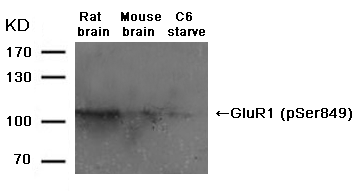
蛋白免疫印迹(WB)
背景介绍
蛋白免疫印迹( Western Blot)是将PAGE(聚丙烯酰胺凝胶电泳)分离的蛋白质样品,转移到固相载体(例如硝酸纤维素薄膜)上,固相载体以非共价键形式吸附蛋白质,且能保持电泳分离的多肽类型及其生物学活性不变。以固相载体上的蛋白质或多肽作为抗原,与对应的抗体起免疫反应,再与酶或同位素标记的第二抗体起反应,经过底物显色或放射自显影以检测电泳分离的特异性目的基因表达的蛋白成分。
一. 仪器与材料
1. 仪器
1) 电泳仪 2)电转仪 3)摇床 4)化学发光成像仪
2. 材料
1) 30%丙烯酰胺溶液
2) 1.5 mol/L Tris (pH8.8)
3) 1.0 mol/L Tris (pH6.8)
4) 10%SDS
5) 10%过硫酸胺
6) TEMED
7) 5× 上样缓冲液
8) 5× SDS电泳缓冲液
9) 10×转移缓冲液
10) NC膜
11) 洗涤液:TBST(1×TBS,0.1%TWEEN20)
12) 封闭液,抗体稀释液:5%脱脂奶粉的TBST
13) HRP
-羊抗兔IgG
14) ECL底物液A
15) ECL底物液B
二. 操作步骤
1. 组装制胶平台
玻璃板用自来水清洗干净,晾干。去离子水检漏后,用滤纸吸去玻璃板中残留的水。
2. 配制所需浓度分离胶(配方见附表)
注意:如果发现10%SDS有沉淀,可先放置50℃左右的温箱或温水浴中至沉淀溶解,待其冷却后方能配胶; 胶液配好后要充分混匀。
3. 灌分离胶
灌胶时要防止气泡产生,每次不要把枪头中液体完全打尽,以免带进气泡,一块0.75mm厚度的胶需加入约4ml的分离胶,灌完后在胶面上轻轻加入2ml蒸馏水封胶。室温凝胶30-40分钟。胶凝后将封胶水倒掉,并用滤纸吸干微量剩余的水。
4. 配制 5% 浓缩胶(配方见附表)
5. 灌浓缩胶
将浓缩胶加满至整个制胶平板,插上合适的梳子。室温凝胶30分钟后。将玻璃板取出,并将玻璃板反转置于内侧。在电泳缓冲液中将梳子拔出。
6. 样品准备
同时进行样品处理 (样品与5×上样缓冲液按照体积比2: 1混合),100℃煮沸5分钟。将处理好的样品放置4℃冰箱预冷5分钟。如有沉淀,则将样品离心弃沉淀。
7. 上样
在电泳芯内层玻板之间灌满电泳缓冲液;依次点上样品及Marker。
8. 电泳:90V恒压跑浓缩胶,然后120 V恒压跑分离胶
9. 转膜准备
剪好大小与胶相同的1张NC膜和4层滤纸,将膜取出(要用镊子,不可以用手直接接触膜表面)先在水中浸泡10分钟,以除去气泡;然后置于1×转移缓冲液中平衡20分钟。再将滤纸于1×转移缓冲液中浸透平衡,时间不宜过长。按照滤纸-凝胶-膜-滤纸三明治式放好。胶和膜之间不能有气泡。膜在正极,胶在负极。
10. 转膜
检测蛋白分子量
转膜条件
>200kd
200mA过夜,(70V)
100-200kd
150 mA过夜,(60V)或200mA,1.5h~2h
30-100kd
200mA,1h-1.5h,(70V)或100mA过夜(35V)
<30kd
200mA,45min-1h(70V)
将预冷的转移缓冲液加入到转印槽中,按所需条件调好仪器参数,在放置了冰块的水中进行转膜。
11. 收膜和封闭
转膜终止,取出NC膜并用TBST清洗,做好标记后可根据需要置于立春红染液中观察转膜效果,观察完用TBST清洗,然后放入加有封闭液的容器中,室温封闭1 hr左右。封闭后用TBST清洗,或晾干备用保存至-20℃冰箱。
12. 切膜
将转好的膜用TBST先浸泡透,取出后将转有蛋白的一面朝上放置在平板上,用刀按方案所需将膜裁切,并在无蛋白位置作好标记(包括抗体编号,膜编号等),分别放入到各反应槽中。
(磷酸酶实验CIP)
用水稀释10x 磷酸酶buffer至1x工作液,以每个泳道50单位CIP配5ml工作液的体系孵膜,37℃孵
育2小时。将孵完CIP的膜在TBST中洗净。
13. 孵育一抗
将各个反应槽中加入所需反应体积的5%脱脂奶粉TBST,然后按照设计方案的稀释度分别加入相应体积的一抗样品。4℃缓慢摇晃过夜。
14. 洗涤
在各个反应槽中加入适量的TBST,摇床摇动5分钟,然后将TBST倒掉,重复3次。
15. 孵育二抗
配制二抗孵育液:将HRP-羊抗兔IgG二抗按照工作稀释比例加到5%脱脂奶粉TBST中,摇床混匀5分钟。将洗涤过的NC膜全部取出,转移到二抗孵育液中室温1小时。
16. 洗涤
倒掉二抗孵育液,加入适量TBST,摇床摇动5分钟,然后将TBST倒掉,重复3次。
17. 底物反应
配制ECL反应底物:临用前,将ECL底物液A 和ECL底物液B等体积混合并混匀,避光保存备用。
将配好的反应底物液均匀的滴加在膜上,保证所有膜都被反应液完全覆盖后,反应1-2分钟,注意膜的位置不要有气泡。
18. 曝光
预先打开凝胶成像仪,调节至最佳条件,将放有膜的平板放入暗箱内,开始曝光。根据情况选择最适的曝光时间。
19. 结果保存
选择最佳图片,整理归档。
-
WB常见问题分析
WB简介
蛋白印迹是通过聚丙烯酰胺凝胶电泳,将不同分子量大小的蛋白分离并转移到杂交膜上,再通过一抗/二抗复合物对蛋白质进行检测的一种免疫生化技术。
提取标本蛋白并定量
凝胶电泳分离蛋白
转移蛋白质到杂交膜
收膜和封闭
孵育一抗
洗涤
孵育酶联物(二抗或者亲和素-HRP/AP)
洗涤
底物显色或曝光检测
结果分析
1.高背景
可能原因
验证或解决办法
膜污染
使用干净镊子,戴手套操作,避免污染
膜干燥
保证充分的反应液,避免出现干膜现象
封闭不完全
封闭液不合适
延长封闭时间
比较尝试不同封闭液
缓冲液污染
使用新配置的缓冲液
漂洗不完全
增加漂洗时间和缓冲液体积
抗体浓度过高
降低一抗、二抗浓度
抗体与阻断蛋白有交叉反应
选择无交叉反应的封闭液,洗涤液中加入Tween-20可以减少交叉反应
2.信号弱或无信号
可能原因
验证或解决办法
抗原量不足
增加上样量
蛋白质降解
重新制备样品
抗原被封闭液遮挡
优化封闭液,缩短封闭时间,减小封闭液中的蛋白浓度
样本中不含靶蛋白或靶蛋白含量太低
设置阳性对照。若靶蛋白含量低,可适当增加样本上样量
膜的错误选择
选择合适孔径的膜。>22KD 0.45um
<22KD 0.22um
转膜不完全
转膜后确定转膜效率,保证胶与膜充分结合,保证电极正确装配,控制转膜温度,优化转膜电流及时间
甲醇浓度过高
过高的甲醇浓度会导致蛋白质与SDS分离,从而沉淀在凝胶中。同时会使凝胶收缩或变硬,抑制高分子蛋白的转移。因此,要根据不同分子量选择合适的甲醇浓度
洗膜过度
缩短洗涤时间或减少洗涤次数
封闭过度
减少封闭剂的量或缩短封闭时间。更换不同封闭剂类型
一抗失效
选择在有效期内的抗体,抗体避免反复冻融。选择现配现用的工作液
一抗反应不充分
增加抗体浓度,延长孵育时间
试剂之间不匹配
一抗与组织种属,一抗与二抗或和底物与酶系统之间不匹配。通过设置内参可以验证二级检测系统的有效性
HRP 抑制
所有溶液和容器中避免含有叠氮化钠
酶和底物失效
直接将酶和底物进行混合,如果不显色说明酶失活了。选择在有效期内、有活性的酶联物,使用新鲜底物
曝光时间过短
延长曝光时间
3.非特异性条带
可能原因
验证或解决办法
蛋白上样量过大
降低样本上样量
蛋白降解
使用新鲜制备的样本,并使用蛋白抑制剂
细胞传代过多,导致蛋白变异
使用原代或传代少的细胞做对照
抗体浓度过高
降低抗体浓度
交叉反应
选择单克隆抗体或亲和纯化的抗体,保证抗体特异性
不同异构体的存在
有些来源于同一基因的蛋白有不同异构体,每个异构体蛋白大小都是不同的
二抗非特异性结合
增加二抗对照,选择其他二抗
底物太灵敏
选择合适底物
曝光时间过长
减少曝光时间
4. 条带位置不对
可能原因
验证或解决办法
二聚体或多聚体的存在
增加蛋白质变性过程及强度
蛋白修饰
如糖基化、磷酸化等修饰状态会导致蛋白分子量增加
5. 条带内出现不显影圆点
可能原因
验证或解决办法
转膜过程有气泡
转膜时赶尽气泡
6. 条带不完整
可能原因
验证或解决办法
底物孵育不均匀
均匀孵育底物
7. 微笑条带
可能原因
验证或解决办法
电泳速度过快
减少电压等减慢电泳速度
电泳温度过高
在冷室或冰浴中进行电泳
8. 反影(白色条带,黑色背景)
可能原因
验证或解决办法
HRP浓度过高
降低二抗浓度
9. 背景有黑色斑点
可能原因
验证或解决办法
转膜时有气泡或抗体分布不均匀
尽量去除气泡,抗体孵育时保持摇动
封闭剂中有聚集体
使用前过滤封闭试剂
抗体与封闭剂反应
选择合适的封闭剂
HRP耦联二抗中有聚集体
过滤二抗试剂,去除聚集体
10.Markera变黑
可能原因
验证或解决办法
抗体与marker蛋白反应
在marker和样本之间空出一个孔不上样
WB实验的主要试剂
1.WB实验膜
PVDF膜,硝酸纤维素膜或尼龙膜。
2.封闭试剂
BSA或脱脂奶粉。
3.蛋白Marker
主要目的是确定靶蛋白的分子量大小;使用预染Marker还可以实时检测电泳分离情况并可以转移到膜上。
4.蛋白提取及定量
类别
品名
SAB对应货号
蛋白提取
总蛋白提取试剂盒
核蛋白提取试剂盒
膜蛋白提取试剂盒
蛋白定量
BCA 蛋白定量试剂盒
Bradford 蛋白定量试剂盒
5.一抗
*选择适合检测标本种属的一抗(说明书上有验证信息)
*选择适用于WB实验方法的一抗(说明书上有验证信息)
*选择单克隆抗体或经过亲和纯化的多克隆抗体
*根据说明书推荐的浓度优化最佳的抗体稀释比
6.内参
内参的作用:
1).检测整个WB实验过程及体系是否正常工作。
2).半定量的标准。
内参的选择:
一般为稳定表达的管家基因蛋白,来源最好选择和同时使用的一抗来源相同的,以方便二抗的选择。
内参选择:
MW(kDa) 全细胞 线粒体 细胞核 细胞膜 Vinculin(124kDa) Alzheimer pathway
.jpg)
免疫组化疑难解答
Troubleshooting tips for IHC common problems:
1. Non-specifc staining
2. No staining
3. Weak staining
4. Strong Staining
1.Non-specifc staining
Causes Solutions Improper preparation of sections Improve ways of sampling and preparation Inadequate deparaffinization of the sections Increase the deparaffinization time Tissue contains endogenous peroxidase Use 0.3% v/v fresh H2O2 for blocking and increase the blocking incubation time Tissue contains endogenous biotin Use IHC biotin blocking agent Blocking of protein may be insufficient Increase the blocking time Charge adsorption Block with nonimmune animal serum The antibody is not pure Change suitable antibody Primary antibody concentration may be too high Try decreasing the antibody concentration The sections have dried out Avoid sections being dried out in the process of experiments Washes may be insufficient Increase the times of washes and the washing time Non-specifc staining: Improved: 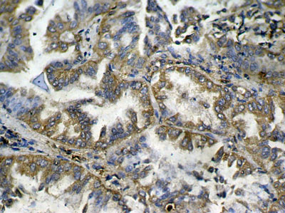
Immunohistochemical analysis of paraffin-embedded human lung carcinoma tissue showing cytoplasmic and nuclear staining using NFκB-p65 Phospho-Ser276 Antibody #11011.
2.No staining
Causes Solutions Improper tissue processing Try to improve the condition and sampling again No antigen in the tissue Set a positive control to verify the experiment results The antibody is not active Don’t use out-of-date antibody kits Incompatible secondary and primary antibodies Use secondary antibody that was raised against the species in which the primary was raised Incompatible staining system Change compatible staining system Improper operation and leave out important steps Follow strict operating procedure and set a positive control No staining: Improved: 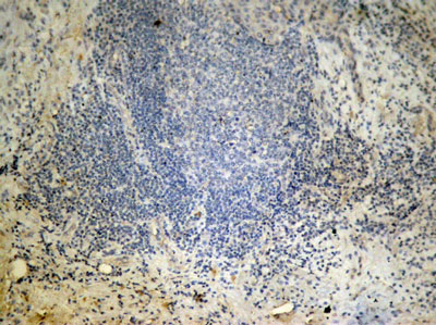
Immunohistochemical analysis of paraffin-embedded human breast carcinoma tissue using Histone H3 Di-Methyl-Lys27 Antibody #11583.
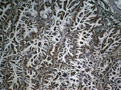
3.Weak staining
Causes Solutions Improper tissue fixation or too high temperature when fixing Use appropriate fixation way or fixation time Too high baking slides temperature and too long baking time Choose appropriate temperature and time for baking slides The antigen may be damaged Let fresh tissues be fixed in time and for not to exceed 24 hours Over blocking of protein Reduce the blocking time The antibody has drained away Ensure that the sections are placed in a horizontal position when incubating The antibody concentration may be too low or incubation time may be too short Increase the antibody concentration and incubation time The room temperature may be too low. Lower than 15℃. Incubator at 37℃ or increase incubation time No draining off buffer solution when adding the reagent results in the reagent being diluted. Do drain off buffer solution but avoid sections being dried out. Excessive washing Wash moderately Always verify the expiration date of the reagent prior to use Change reagents timely Weak staining: Improved: 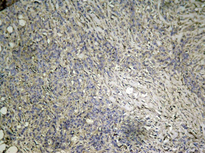

Immunohistochemical analysis of paraffin-embedded human breast carcinoma tissue using mTOR Phospho-Ser2448 Antibody#11221.
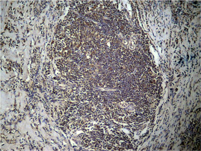
4.Strong Staining
Causes Solutions The primary antibody concentration may be too high or incubation time may be too long Reduce the primary antibody concentration or incubation time. The incubation temperature may be too high Incubate at 4℃ or at room temperature The incubation time of HRP conjugated secondary antibody may be too long Reduce the incubation time Inadequate washing Increase the times of washing Strong Staining: Improved: .jpg)
.jpg)
Immunohistochemical analysis of paraffin-embedded human breast carcinoma tissue using FKHR Phospho-Ser256 Antibody#11115.
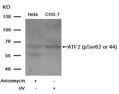
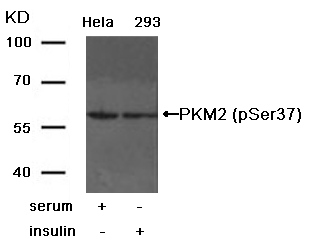
免疫印迹疑难解答
Troubleshooting tips for western blotting common problems:1.High background2.Low or no signal3.Non-specific bands4.Wrong band location5.Invisible dots on the bands6.Incomplete bands7.Smile effect of the bands8.White bands on a black blot9.Black dots on the blot10.Marker lane is black1.High Background
Causes Solutions Membrane fouling Use clean tweezer and operate with gloves to prevent membrane fouling. The membrane has dried out Incubate in sufficient reaction solution to prevent the membrane from drying out. Blocking insufficient Increase the blocking incubation time Inappropriate blocking buffer Switch different blocking buffer Buffer solution has been contaminated Use new buffer solution Incomplete washing Increase washing time and washing buffer’s volume The antibody concentration may be too high Decrease the concentration of primary antibody or secondary antibody Cross-reaction between antibody and blocking agent Choose blocking solution without cross-reaction. Add Tween-20 to the washing buffer to reduce cross reaction. High background: 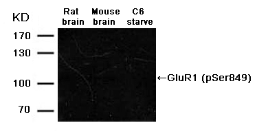
Improved: .gif)
2. Low or no signal
Causes solutions Insufficient antigen Increase amount of loading samples Protein degradation Re-prepare samples The antigen is blocked by blocking buffer Optimize blocking solution, decrease blocking time or decrease the concentration of proteins in the blocking solution. No or low level of target protein in samples Run a positive control. If the level of target protein in samples is low, try to increase amount of loading sample. Wrong choice of membrane Choose suitable pore size membrane. Use 0.45um size membrane for proteins larger than 22KD. Use 0.2 µm size membrane for proteins smaller than 22 KD. Poor transfer of protein to membrane Make sure there are no air bubbles between the gel and membrane during transfer. Always ensure assembling electrode correctly. Control transfer temperature and optimize transfer electricity and time. Methanol concentration may be too high. Too high concentration of methanol may result in the separation of protein and SDS and thus cause protein precipitation in the gel. At the same time, it may cause shrinking or hardening of the gel to inhibit transferring of high molecular weight proteins. As a result, choose suitable methanol concentration according to different molecular weight. Excessive washing of membrane Reduce washing time and washing times Over blocking Lower the concentration of your blocking solution and shorten blocking time. Change blocking solutions. The primary antibody is inactive Use effective antibody in expiration, avoid freezing- thawing repeatedly, and use fresh solution. Insufficient reaction of antibody to membrane Increase the concentration of the antibody and the incubation time. The reagents are not compatible with each other Primary antibody and species, primary antibody and secondary antibody, or enzyme and substrate are not compatible. Setting loading control can validate the secondary detecting system. HRP Inhibited Avoid sodium azide in all solutions and containers Enzyme or substrate is inactive. Directly mix enzyme and substrate. If no color, the enzyme doesn’t work. Choose active conjugated reagent. Use fresh substrate. Exposure time is too short Increase the exposure time Low or no signal Improved 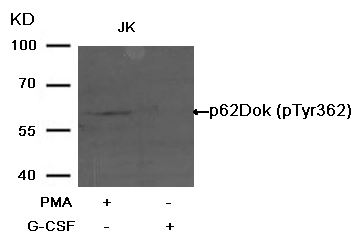
3. Non-specific bands
Causes Solutions Amount of loading samples is too large Decrease amount of loading samples Protein degradation Use fresh samples and use protein inhibitor Cells were cultured too many passages to result in protein variation Use primary cells or less passaging cells to run a control Antibody Concentration is too high Decrease the concentration of the primary or secondary antibody Cross-reaction Choose monoclonal antibody or affinity purified antibody to ensure antibody specificity Existence of the different protein isoforms Some proteins derived from the same gene have different isoforms. The size of every isoform protein is different. Non-specific signal caused by the secondary antibody Run a secondary antibody control or choose other secondary antibody Substrate is too sensitive Use suitable substrate Exposure time is too long Reduce the exposure time Non-specific bands: Improved: 
4.Wrong band location
Causes Solutions Existence of dimer or polymer Increase a process or intensity of protein denaturation Modification of proteins Modification of proteins, such as glycosylation or phosphorylation, can result in an increase of molecular weight of protein. 5.Invisible dots on the bands
Causes Solutions Air bubbles were trapped in the gap of gel and membrane during transferring. Remove bubbles in the gap of gel and membrane when preparing for transferring. Invisible dots on the bands: Improved: 
6.Incomplete bands
Causes Solutions Substrate is not well-distributed during incubation Even the substrate during incubation Incomplete bands: Improved: 
7.Smile effect of the bands
Causes Solutions Migration was too fast during electrophoresis Reduce the voltage to slow down the migration Migration was too hot Run the gel in the cold room or in ice Smile effect of the bands: Improved: 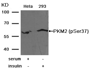
8.White bands on a black blot
Causes Solutions HRP concentration is too high Decrease the secondary antibody concentration White bands on a black blot: Improved: 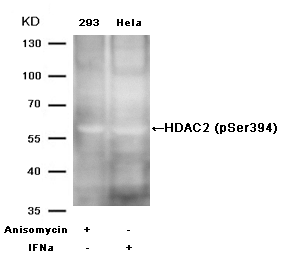
9.Black dots on the blot
Causes Solutions Air bubbles were trapped against the membrane during transferring or the antibody is not well distributed during incubation Try to remove bubbles. Keep shaking when incubating the antibody. The blocking agent was not well dissolved. Filter the blocking agent. The antibody reacts with the blocking solution. Choose suitable blocking solution There are aggregates in the HRP conjugated secondary antibody. Filter the secondary antibody agent and remove the aggregates. Black dots on the blot: Improved: 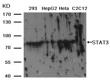

10.Marker lane is black
Causes Solutions The antibody reacts with the MW marker Add a blank lane between the marker and the adjacent sample lane
